alb3770012
Breast Anatomy, Illustration
| Teilen |
|---|
 Pinterest Pinterest |
 Twitter Twitter |
 Facebook Facebook |
 Link kopieren Link kopieren |
 Email Email |
|
Zu einem anderen Lightbox hinzufügen |
|
Zu einem anderen Lightbox hinzufügen |


Haben Sie bereits ein Konto? Anmelden
Sie haben kein Konto? Registrieren
Dieses Bild kaufen.
Nutzung auswählen:

Titel: Breast Anatomy, Illustration
Untertitel: Siehe automatische Übersetzung
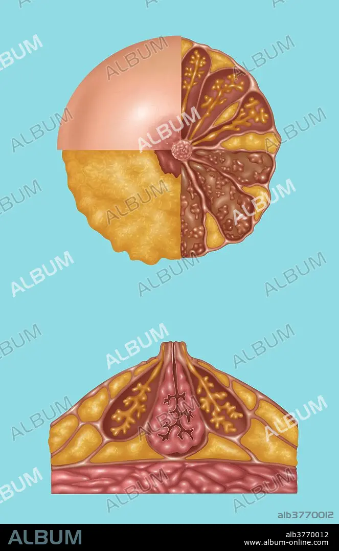
Illustration detailing the anatomy of a female breast from a front view (top) and a side view (bottom). The top image is in three sections. The areola, montgomery's tubercules, and nipple are in the light pink section. The nipple and subareolar musculature and subcutaneous fat are in the orange section. On the right half is: mammary fat (orange), lactiferous ducts (orange stems), acini (end of orange stems), ampulla (on orange stem), coopers ligaments (light pink strands), lobe and lobules (brown outlines and pink groups inside the brown), and interlobular connective tissue (brown area). The bottom image shows montgomery's gland and superficial fascia (outer lining), subcutaneous fat (orange outer sections), ampulla and lactiferous duct and connective tissue (pink center), coopers ligaments, mammary fat (orange bottom sections), pectoral fascia, and pectoralis major (pink at bottom).
Illustration detailing the anatomy of a female breast from a front view (top) and a side view (bottom). The top image is in three sections. The areola, montgomery's tubercules, and nipple are in the light pink section. The nipple and subareolar musculature and subcutaneous fat are in the orange section. On the right half is: mammary fat (orange), lactiferous ducts (orange stems), acini (end of orange stems), ampulla (on orange stem), coopers ligaments (light pink strands), lobe and lobules (brown outlines and pink groups inside the brown), and interlobular connective tissue (brown area). The bottom image shows montgomery's gland and superficial fascia (outer lining), subcutaneous fat (orange outer sections), ampulla and lactiferous duct and connective tissue (pink center), coopers ligaments, mammary fat (orange bottom sections), pectoral fascia, and pectoralis major (pink at bottom).
Bildnachweis: Album / Science Source / Gwen Shockey
Freigaben (Releases): ? Modellfreigabe: Nein - ? Eigentumsfreigabe: Nein
Rechtefragen?
Rechtefragen?
Bildgröße: 3996 × 6168 px | 70.5 MB
Druckgröße: 33.8 × 52.2 cm | 1573.2 × 2428.3 in (300 dpi)
Schlüsselwörter: ANATOMIE • ILLUSTRATION • ILLUSTRATIONS
