alb3793880
Historical Illustration of Honey Bee Eye
| Teilen |
|---|
 Pinterest Pinterest |
 Twitter Twitter |
 Facebook Facebook |
 Link kopieren Link kopieren |
 Email Email |
|
Zu einem anderen Lightbox hinzufügen |
|
Zu einem anderen Lightbox hinzufügen |


Haben Sie bereits ein Konto? Anmelden
Sie haben kein Konto? Registrieren
Dieses Bild kaufen

Titel:
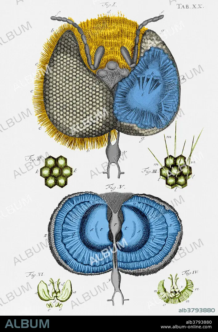
Historical Illustration of Honey Bee Eye
Untertitel:
Siehe automatische Übersetzung
Honey bee eye. Drawing of the structure of the eye of a honey bee (Apis mellifera) by the Dutch naturalist Jan Swammerdam (1637-1680). The individual components of the eye are shown. These consist of long rods (ommatidia) which have a hexagonal lens at the top end, and taper to a nerve bundle at the base. Swammerdam studied medicine, but never practiced it, and instead used his microscope to study insects. This, and hundreds of other drawings, were published posthumously in the celebrated Bybel der Nature in 1737. His studies of metamorphosis form the basis of insect classification today. He also studied mammalian muscles, and discovered red blood cells.
Bildnachweis:
Album / Science Source / Photo Researchers
Freigaben (Releases):
Model: Nein - Eigentum: Nein
Rechtefragen?
Rechtefragen?
Bildgröße:
3322 x 4809 px | 45.7 MB
Druckgröße:
28.1 x 40.7 cm | 11.1 x 16.0 in (300 dpi)
Schlüsselwörter:
ABDRUCK • ANATOMIE • AUGE • AUGE, SEHEN • AUGEN • DIAGRAMM • FARBE • FARBIG • FAUNA • HONIG • ILLUSTRATION • ILLUSTRATIONS • INSEKT • INSEKTEN • ZOOLOGIE
