alb13913848
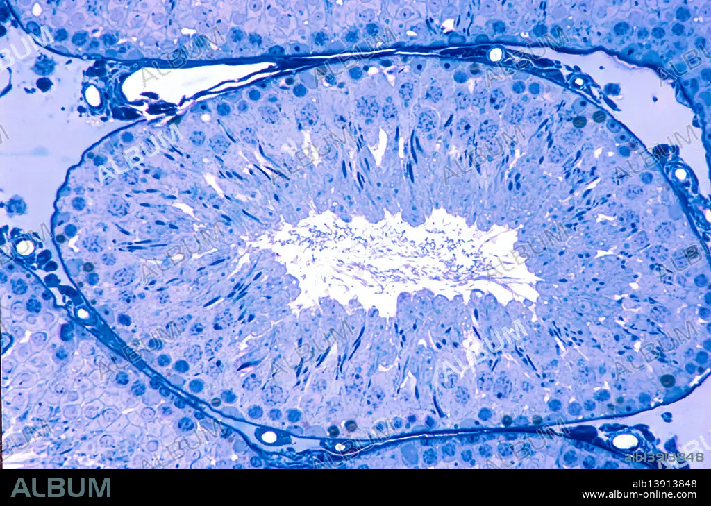
Rat testis, light micrograph
| Teilen |
|---|
 Pinterest Pinterest |
 Twitter Twitter |
 Facebook Facebook |
 Link kopieren Link kopieren |
 Email Email |
|
Zu einem anderen Lightbox hinzufügen |
|
Zu einem anderen Lightbox hinzufügen |


Haben Sie bereits ein Konto? Anmelden
Sie haben kein Konto? Registrieren
Dieses Bild kaufen

Titel:
Rat testis, light micrograph
Untertitel:
Siehe automatische Übersetzung
Rat testis, light micrograph. Cross-section of a seminiferous tubule, lined by a seminiferous epithelium where spermatogonia and Sertoli cells are located at the base, primary spermatocytes in pachytene of prophase I of the first meiotic division occupy an intermediate position, and spermatids with elongated nuclei are found near the lumen. 0.5 micrometre thick section of plastic embedded material stained with toluidine blue.
Bildnachweis:
Album / JOSE CALVO / SCIENCE PHOTO LIBRARY
Freigaben (Releases):
Model: Nein - Eigentum: Nein
Rechtefragen?
Rechtefragen?
Bildgröße:
4800 x 3168 px | 43.5 MB
Druckgröße:
40.6 x 26.8 cm | 16.0 x 10.6 in (300 dpi)
