alb3886157
Microscopic view of a blastula during pregnancy.
| Teilen |
|---|
 Pinterest Pinterest |
 Twitter Twitter |
 Facebook Facebook |
 Link kopieren Link kopieren |
 Email Email |
|
Zu einem anderen Lightbox hinzufügen |
|
Zu einem anderen Lightbox hinzufügen |


Haben Sie bereits ein Konto? Anmelden
Sie haben kein Konto? Registrieren
Dieses Bild kaufen

Titel:
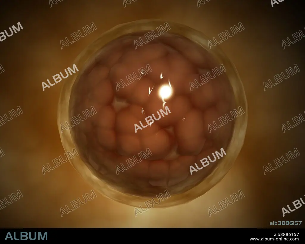
Microscopic view of a blastula during pregnancy.
Untertitel:
Siehe automatische Übersetzung
Microscopic view of a blastula during pregnancy. After the cleavage has produced over 100 cells, the embryo is called a blastula. The blastula is usually a spherical layer of cells (the blastoderm) surrounding a fluid-filled or yolk-filled cavity (the blastocoel).
Bildnachweis:
Album / Stocktrek Images
Freigaben (Releases):
Model: Nein - Eigentum: Nein
Rechtefragen?
Rechtefragen?
Bildgröße:
4920 x 3690 px | 51.9 MB
Druckgröße:
41.7 x 31.2 cm | 16.4 x 12.3 in (300 dpi)
Schlüsselwörter:
DURCHSCHEINEND • EMBRYO • FOETUS • FOTUS • FÖTUS • ILLUSTRATION • ILLUSTRATIONS • MIKROBIOLOGIE • MOLEKULARBIOLOGIE • NAHAUFNAHME • SCHWANGERSCHAFT • VERGROESSERUNG • VERGRÖSSERUNG • ZYTOLOGIE
