alb3794817
ear canal
| Teilen |
|---|
 Pinterest Pinterest |
 Twitter Twitter |
 Facebook Facebook |
 Link kopieren Link kopieren |
 Email Email |
|
Zu einem anderen Lightbox hinzufügen |
|
Zu einem anderen Lightbox hinzufügen |


Haben Sie bereits ein Konto? Anmelden
Sie haben kein Konto? Registrieren
Dieses Bild kaufen.
Nutzung auswählen:

Titel: ear canal
Untertitel: Siehe automatische Übersetzung
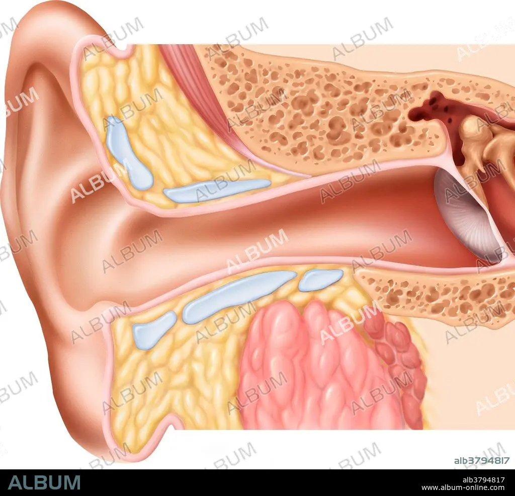
Illustration of the normal ear canal. The inner ear contains a maze of fluid-filled passages called the labyrinth. The cochlea is a hollow spiral containing microscopic hairs that respond to sound vibrations conveyed from the middle ear. The near end of the spiral picks up high-frequency sound; the central (far) end detects low frequency sound. Nerves connected to the cochlea carry sound impulses to the brain. The semi-circular canals provide the sense of balance by detecting movement of fluid through the three planes.
Illustration of the normal ear canal. The inner ear contains a maze of fluid-filled passages called the labyrinth. The cochlea is a hollow spiral containing microscopic hairs that respond to sound vibrations conveyed from the middle ear. The near end of the spiral picks up high-frequency sound; the central (far) end detects low frequency sound. Nerves connected to the cochlea carry sound impulses to the brain. The semi-circular canals provide the sense of balance by detecting movement of fluid through the three planes.
Kategorie: WISSENSCHAFT
Bildnachweis: Album / Science Source / Brian Evans
Freigaben (Releases): ? Modellfreigabe: Nein - ? Eigentumsfreigabe: Nein
Rechtefragen?
Rechtefragen?
Bildgröße: 1800 × 1641 px | 8.5 MB
Druckgröße: 15.2 × 13.9 cm | 708.7 × 646.1 in (300 dpi)
Schlüsselwörter: ILLUSTRATION • ILLUSTRATIONS • OHR • WISSENSCHAFT
