alb10686068
Neurofibromatosis type I (NF1), MRI
| Teilen |
|---|
 Pinterest Pinterest |
 Twitter Twitter |
 Facebook Facebook |
 Link kopieren Link kopieren |
 Email Email |
|
Zu einem anderen Lightbox hinzufügen |
|
Zu einem anderen Lightbox hinzufügen |


Haben Sie bereits ein Konto? Anmelden
Sie haben kein Konto? Registrieren
Dieses Bild kaufen.
Nutzung auswählen:

Titel:
Neurofibromatosis type I (NF1), MRI
Untertitel:
Siehe automatische Übersetzung
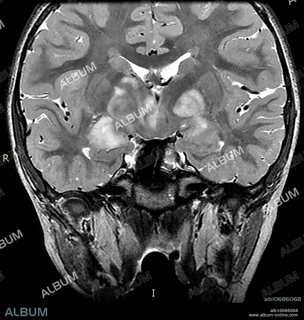
This coronal (from the front) T2 weighted MRI of a 15 year old with known NF1 demonstrates the typical appearance of foci of abnormal increased T2 signal within the basal ganglia, internal capsule and thalamus commonly seen in NF1. These foci usually wax and wane and disappear as the subject approaches the age of 20.
Bildnachweis:
Album / Science Source / Living Art Enterprises
Freigaben (Releases):
Bildgröße:
3900 x 3900 px | 43.5 MB
Druckgröße:
33.0 x 33.0 cm | 13.0 x 13.0 in (300 dpi)
