alb10657099
Lingual Thyroid, CT Scan
| Teilen |
|---|
 Pinterest Pinterest |
 Twitter Twitter |
 Facebook Facebook |
 Link kopieren Link kopieren |
 Email Email |
|
Zu einem anderen Lightbox hinzufügen |
|
Zu einem anderen Lightbox hinzufügen |


Haben Sie bereits ein Konto? Anmelden
Sie haben kein Konto? Registrieren
Dieses Bild kaufen.
Nutzung auswählen:

Titel:
Lingual Thyroid, CT Scan
Untertitel:
Siehe automatische Übersetzung
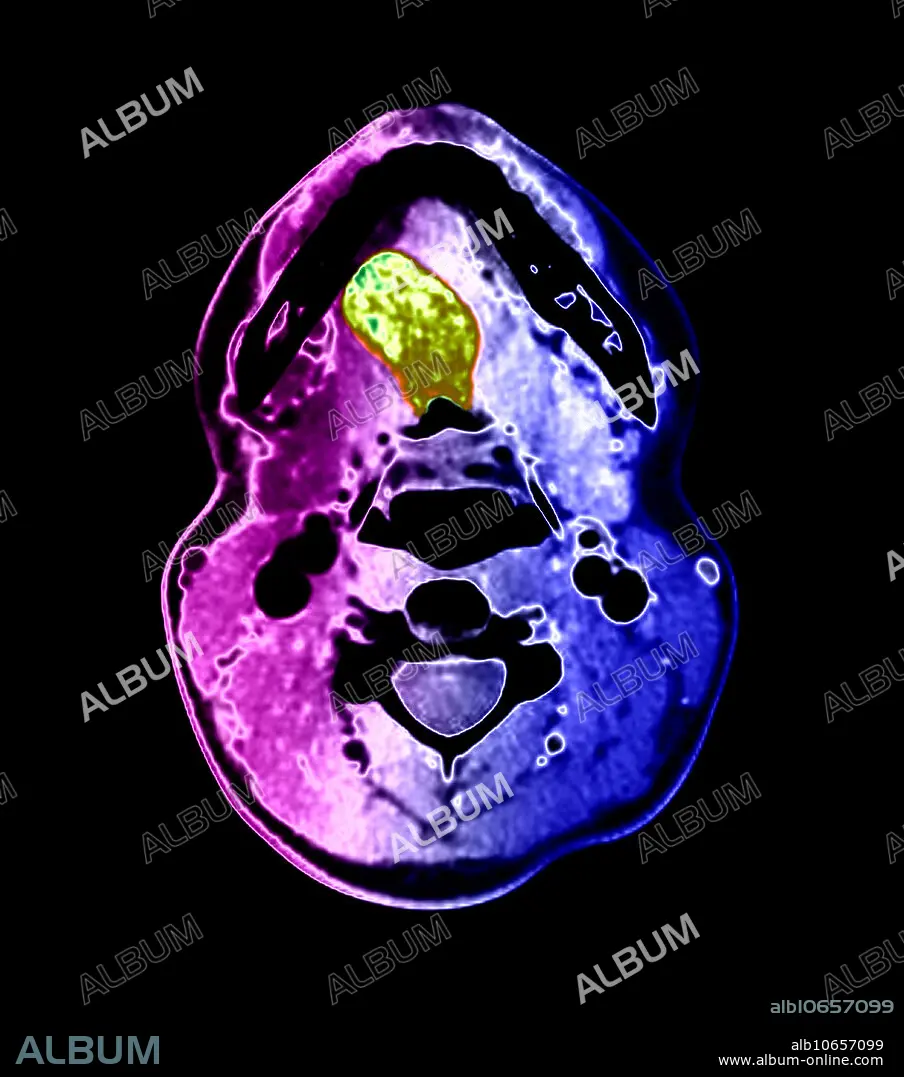
This colour-enhanced, axial (cross-sectional) CT image through the oral cavity demonstrates a large, abnormal density (green) in the tongue. This represents lingual thyroid tissue. This patient had no thyroid tissue in the expected region of the lower neck. The thyroid gland develops near the foramen cecum of the tongue and normally descends along a tract called the thyroglossal duct to the lower neck. Rarely, this normal descent does not occur and thyroid tissue remains in the tongue, but remnants of thyroid tissue and the duct itself may occur anywhere along the tract. Often, however, no thyroid tissue is present in the lower neck, only in the tongue base, as in this case.
Persönlichkeiten:
Bildnachweis:
Album / Science Source / Living Art Enterprises
Freigaben (Releases):
Model: Nein - Eigentum: Nein
Rechtefragen?
Rechtefragen?
Bildgröße:
4200 x 4755 px | 57.1 MB
Druckgröße:
35.6 x 40.3 cm | 14.0 x 15.8 in (300 dpi)
Schlüsselwörter:
