alb3824782
Double Focus X-ray Tube, 1896
| Teilen |
|---|
 Pinterest Pinterest |
 Twitter Twitter |
 Facebook Facebook |
 Link kopieren Link kopieren |
 Email Email |
|
Zu einem anderen Lightbox hinzufügen |
|
Zu einem anderen Lightbox hinzufügen |


Haben Sie bereits ein Konto? Anmelden
Sie haben kein Konto? Registrieren
Dieses Bild kaufen

Titel:
Double Focus X-ray Tube, 1896
Untertitel:
Siehe automatische Übersetzung
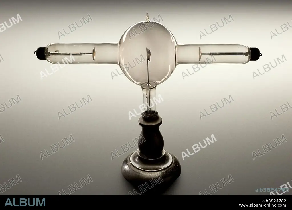
Double focus x-ray tube, Europe, 1896. This tube worked by using an alternating current, which accelerates electrons towards an aluminium plate. This produced x-rays at both ends of the tube. Wilhelm Roentgen, a German physician, took the first x-ray in 1896 of his wife's left hand. Dense areas of bone show up as white whilst soft tissue allow the x-ray to pass through undeterred. Very quickly x-rays proved their usefulness as a diagnostic and therapeutic tool in medicine. Within six months of Roentgen's announcement, x-rays were being used by battlefield physicians to locate bullets in wounded soldiers. X-rays allowed physicians their first look inside the body without resorting to surgery.
Bildnachweis:
Album / Science Source / Wellcome Images
Freigaben (Releases):
Model: Nein - Eigentum: Nein
Rechtefragen?
Rechtefragen?
Bildgröße:
4224 x 2823 px | 34.1 MB
Druckgröße:
35.8 x 23.9 cm | 14.1 x 9.4 in (300 dpi)
Schlüsselwörter:
