alb10673150
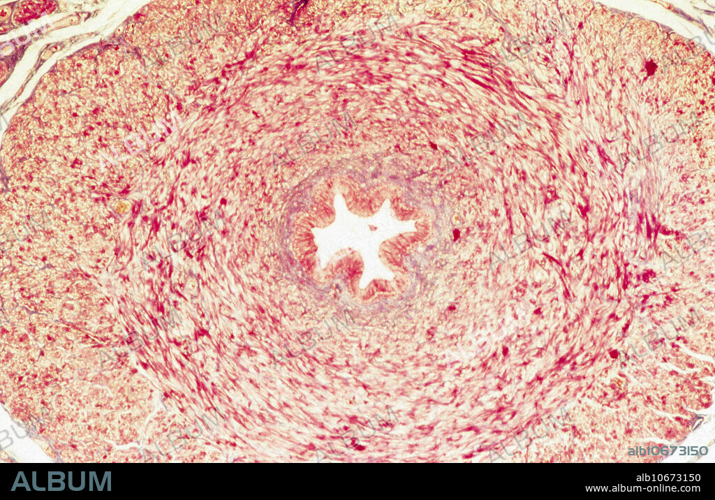
Ureter, LM

|
Add to another lightbox |
|
Add to another lightbox |


Title:
Ureter, LM
Caption:
Histological appearance of a cross section of the ureter. The folded ureter epithelium in the centre is surrounded by a tick layer of smooth muscle (yellow) and fibrous (red) tissue. Elastica van Gieson staining and 20x magnification.
Credit:
Album / Nature's Faces/Science Source
Releases:
Model: No - Property: No
Rights questions?
Rights questions?
Image size:
3232 x 2092 px | 19.3 MB
Print size:
27.4 x 17.7 cm | 10.8 x 7.0 in (300 dpi)
Keywords:
EPITHELIUM • FIBER • FIBRE • HISTOLOGIC • HISTOLOGICAL • HISTOLOGY • LIGHT • LM • MICROGRAPH • MICROGRAPHY • MICROSCOPY • MUSCLE • NORMAL • SMOOTH • STAIN • URETER • URETHER • URINARY DUCT
 Pinterest
 Twitter
 Facebook
 Copy link
 Email
Email

