alb10694504
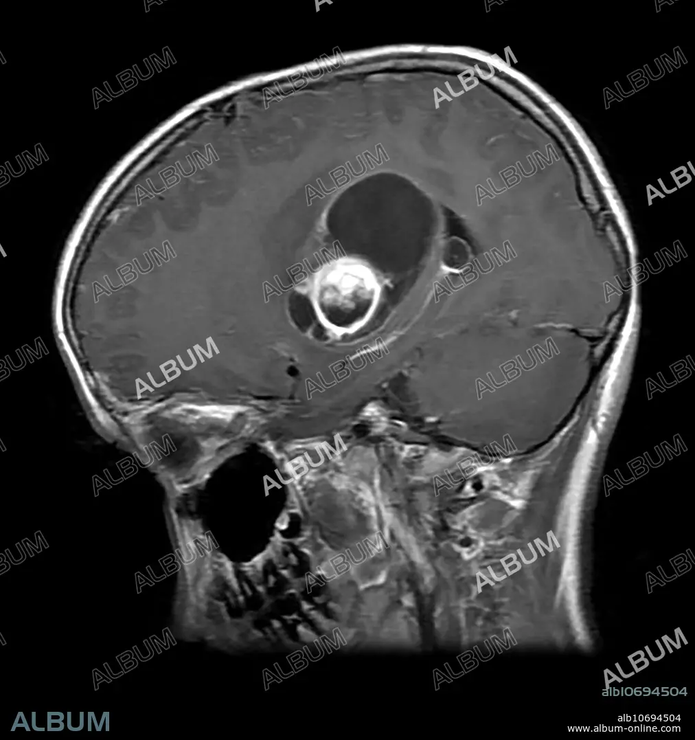
MRI Pilocytic Astrocytoma
| Share |
|---|
 Pinterest Pinterest |
 Twitter Twitter |
 Facebook Facebook |
 Copy link Copy link |
 Email Email |
|
Add to another lightbox |
|
Add to another lightbox |


Title:
MRI Pilocytic Astrocytoma
Caption:
This axial (cross sectional) T1 weighted MR image with contrast shows a partially cystic and solid enhancing mass in the basal ganglia, internal capsule and thalamic regions with associate mass effect in a 25 year old. This represents a WHO grade 1 astrocytoma called a pilocytic astrocytoma.
Personalities:
Credit:
Album / Living Art Enterprises, LLC/Science Source
Releases:
Model: No - Property: No
Rights questions?
Rights questions?
Image size:
3900 x 3947 px | 44.0 MB
Print size:
33.0 x 33.4 cm | 13.0 x 13.2 in (300 dpi)
