alb3785092
Anatomy of Human Tongue, Illustration
| Share |
|---|
 Pinterest Pinterest |
 Twitter Twitter |
 Facebook Facebook |
 Copy link Copy link |
 Email Email |
|
Add to another lightbox |
|
Add to another lightbox |


Buy this image.
Select the use:

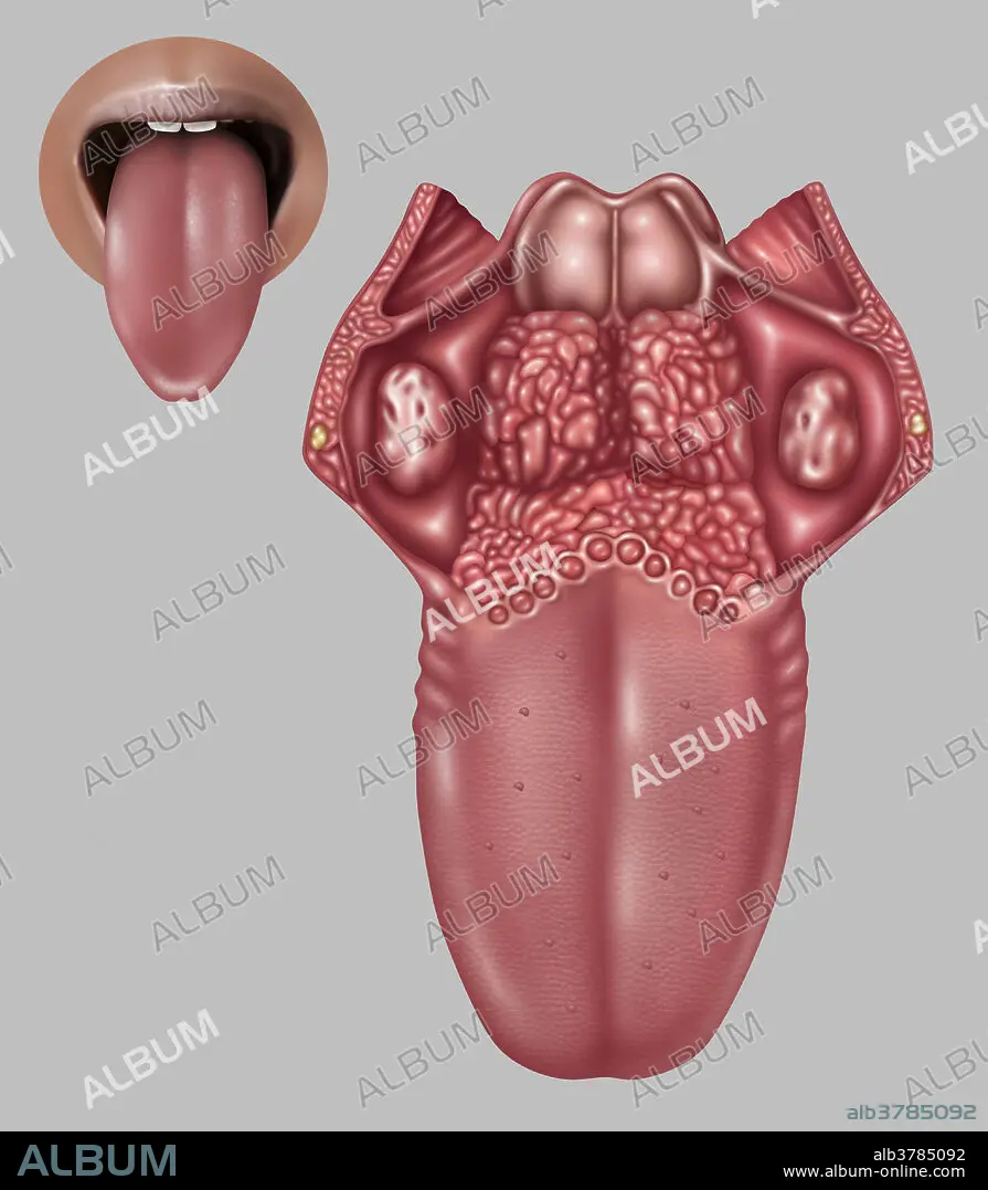
Title: Anatomy of Human Tongue, Illustration
Caption: Illustration depicting the anatomy of the human tongue. Listed at left from top to bottom are: palatopharyngeal arch (at back on sides), lingual tonsil (textured area at back), palatoglossal arch, foliate papillae (feathered part on side of tongue), medial sulcus of the tongue (middle crease), filiform papilla (bottom outer area), epiglottis (top middle), palatine tonsil (round textured areas on upper sides), terminal sulcus (middle centre back), vallate papilla (circumvallate papilla) (round and forming a 'V'), fungiform papilla (middle sides of tongue).
Credit: Album / Science Source / Gwen Shockey
Releases: ? Model Release: No - ? Property Release: No
Rights questions?
Rights questions?
Image size: 3948 × 4515 px | 51.0 MB
Print size: 33.4 × 38.2 cm | 1554.3 × 1777.6 in (300 dpi)
Keywords: ANATOMICAL • ANATOMY • ANNOTATED • ARCH • ART • ARTWORK • BACKGROUND • BUDS • CIRCUMVALLATE • DRAWING • EPIGLOTTIS • FILIFORM • FOLIATE • FUNGIFORM • GRAPHIC • GREY • GROSS ANATOMY • HUMAN • HUMANE • ILLUSTRATION • ILLUSTRATIONS • ILUSTRATION • INDIVIDUAL • INFOGRAPHIC • INFORMATION • INSET • LINGUAL • MEDIAL • MEDICAL • MEDICINAL • MOUTH • OF • PALATINE • PALATOGLOSSAL • PALATOPHARYNGEAL • PAPILLA • PAPILLAE • PERSON • PUNT • SCIENCE • SULCUS • TASTE • TERMINAL • THE • TONGUE • TONSIL • VALLATE
