alb9203422
motor neuron

|
Add to another lightbox |
|
Add to another lightbox |


Buy this image.
Select the use:

Title:
motor neuron
Caption:
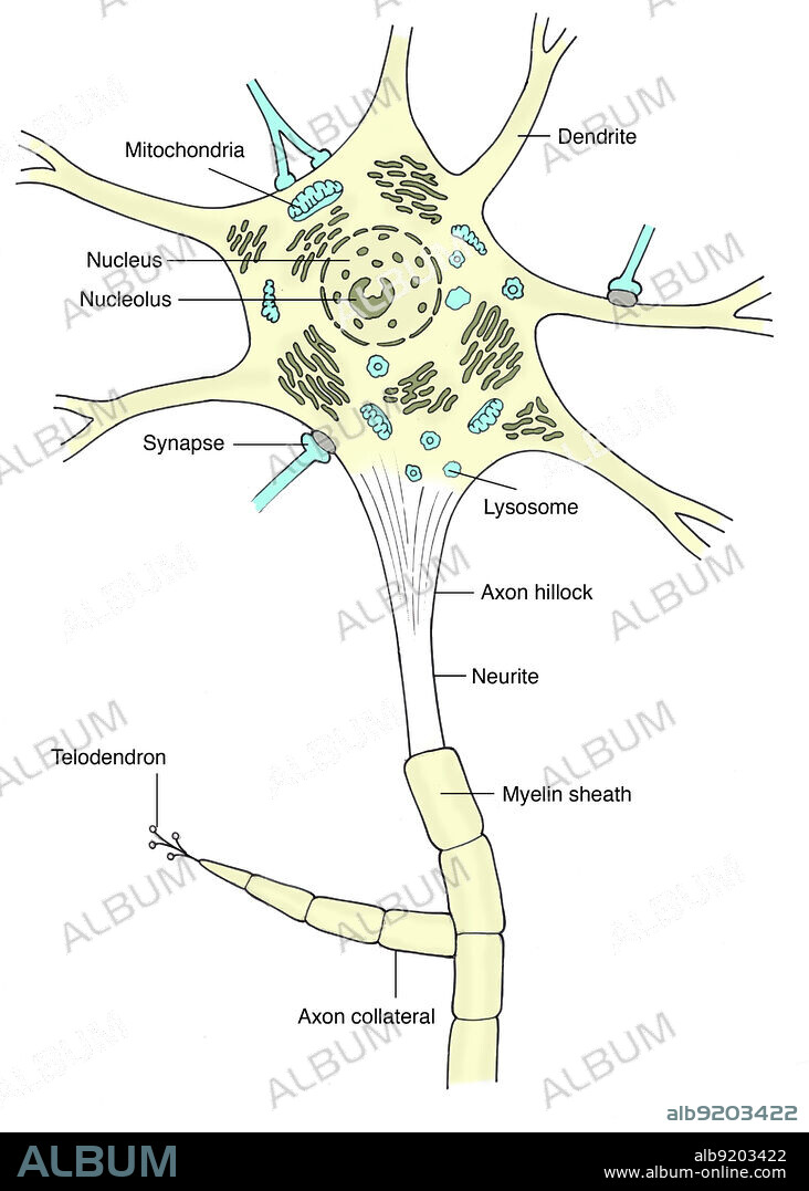
Illustration of a motor neuron, showing mitochondria, dendrites, nucleus, nucleolus, synapses, lysosomes, granular endoplasmic reticulum, axon hillock, neurite, perikaryon (initial segment), myelin sheath, axon collateral, and telodendron. A motor neuron (or motoneuron) is a neuron whose cell body is located in the spinal cord and whose fiber (axon) projects outside the spinal cord to directly or indirectly control effector organs, mainly muscles and glands. Motor neurons' axons are efferent nerve fibers that carry signals from the spinal cord to the effectors to produce effects. Types of motor neurons are alpha motor neurons, beta motor neurons, and gamma motor neurons.
Credit:
Album / Science Source
Releases:
Model: No - Property: No
Rights questions?
Rights questions?
Image size:
Not available
Print size:
Not available
Keywords:
ANATOMY • ART • AXON • BIOLOGY • BODY • CELL • CENTRAL • COLLATERAL • COLORIZED • CORE • CYTOLOGY • CYTON • DENDRITES • DENDRON • ENDOPLASMIC • ENHANCED • ER • EUKARYOTE • FIBERS • FIBRES • GRANULAR • GROSS ANATOMY • HEALTHY • HILLOCK • ILLUSTRATION • ILLUSTRATIONS • LABELED • LYSOSOMES • MEDICAL • MEDICINAL • MITOCHONDRIA • MITOCHONDRIUM • MOTONEURON • MOTOR • MYELIN • NERVE • NERVOUS • NEURITE • NEURON • NEURONAL • NON-PATHOLOGICAL • NORMAL • NUCLEOLUS • NUCLEUS • ORGANELLES • PERIKARYON • PHYSIOLOGIE • PHYSIOLOGY • PROCESS • RETICULUM • RF • SHEATH • SÔMA • SYNAPSE • SYSTEM • TELODENDRIA • TELODENDRON
 Pinterest
 Twitter
 Facebook
 Copy link
 Email
Email
