alb10663034
Casein micelles
| Share |
|---|
 Pinterest Pinterest |
 Twitter Twitter |
 Facebook Facebook |
 Copy link Copy link |
 Email Email |
|
Add to another lightbox |
|
Add to another lightbox |


Buy this image.
Select the use:

Title:
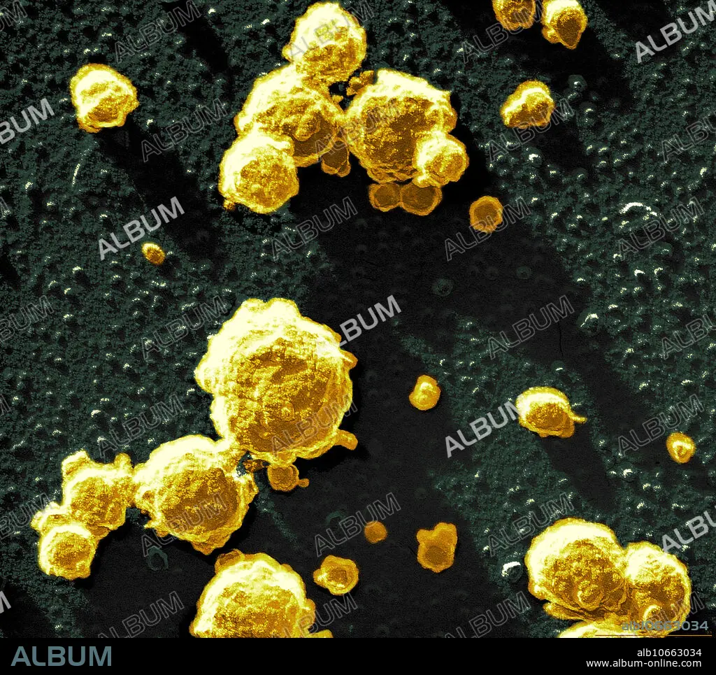
Casein micelles
Caption:
Scanning electron micrograph (SEM) of Casein micelles. Casein micelles are minute protein globules that continually collide and ricochet in fresh milk. Their free path is about 3 micelle diameters. Under the effects of enzymes (chymosis) or acids the proteins coagulate. This image shows the beginning of coagulation, where the casein micelles begin to form clusters. The minute particles on the base are submicellart casein. (Image width: 0.61 micrometer, bar: 0.1 micrometer.)
Credit:
Album / Science Source / SCIMAT
Releases:
Model: No - Property: No
Rights questions?
Rights questions?
Image size:
2688 x 2394 px | 18.4 MB
Print size:
22.8 x 20.3 cm | 9.0 x 8.0 in (300 dpi)
Keywords:
BIOCHEMICAL • BIOCHEMISTRY • CASEIN • CHEMICAL • CHEMISTRY • COAGULATE • COAGULATION • COMPOUND • COMPOUNDS • CURD • CURDS • DAIRY • ELECTRON • GLOBULE • GLOBULES • KITCHENWARE: MILKPOT • MICELLES • MICROGRAPHS • MICROGRAPHY • MILK • MILKPOT • MOLÉCULE • MOLECULES • PRODUCT • PRODUCTS • PROTEIN • PROTEINS • SCANNING • SEM • SEMS • YOGURT • YOGURTS
