alb3772900
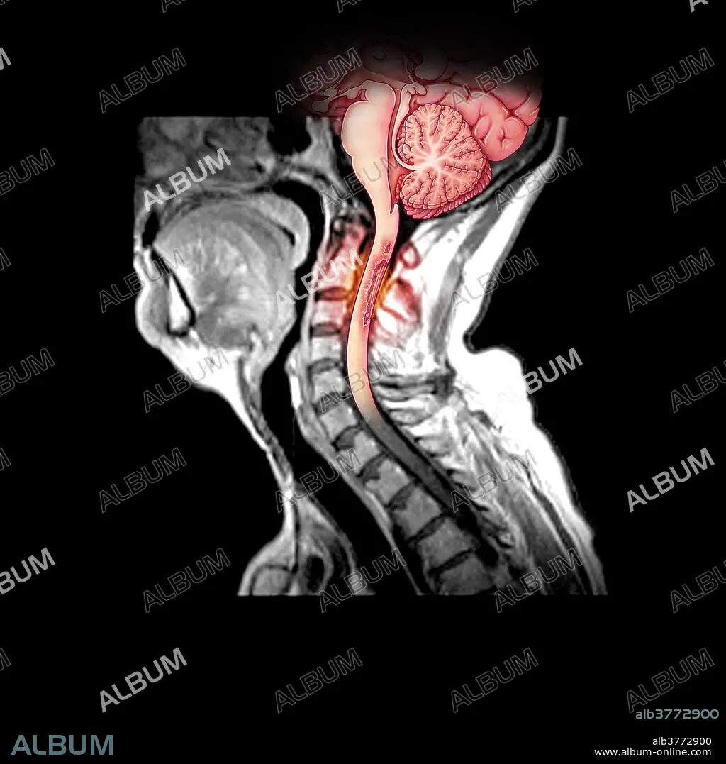
Demyelination of Spinal Cord in MS
| Share |
|---|
 Pinterest Pinterest |
 Twitter Twitter |
 Facebook Facebook |
 Copy link Copy link |
 Email Email |
|
Add to another lightbox |
|
Add to another lightbox |


Buy this image.
Select the use:

Title: Demyelination of Spinal Cord in MS
Caption: This composite illustration of a MRI of the cervical spine/spinal cord shows an area of pathologic enhancement along the posterior (dorsal) aspect of the upper cervical spinal cord. This is in a patient with multiple sclerosis and represents areas of active demyelination.
Credit: Album / Science Source / Evan Oto
Releases: ? Model Release: No - ? Property Release: No
Rights questions?
Rights questions?
Image size: 3600 × 3600 px | 37.1 MB
Print size: 30.5 × 30.5 cm | 1417.3 × 1417.3 in (300 dpi)
Keywords: ART • ARTWORK • BRAIN • CENTRAL • CERVICAL • CONDITION • CORD • DEMYELINATING • DEMYELINATION • DIAGNOSTIC • DISEASE • DISORDER • ENHANCEMENT • ILLUSTRATION • ILLUSTRATIONS • ILUSTRATION • IMAGE • IMAGING • INJURY • LESION • MAGNETIC • MEDICAL • MEDICINAL • MESS • MESSY • MRI • MS • MULTIPLE • NERVOUS • PATHOLOGICAL • PATHOLOGY • PLAQUE • PLATE • RESONANCE • SCLEROSIS • SONORITY • SPINAL • SPINE • SYSTEM • WOUND
