alb10608671
Right Hand Anatomy, Ventral View
| Share |
|---|
 Pinterest Pinterest |
 Twitter Twitter |
 Facebook Facebook |
 Copy link Copy link |
 Email Email |
|
Add to another lightbox |
|
Add to another lightbox |


Buy this image.
Select the use:

Title:
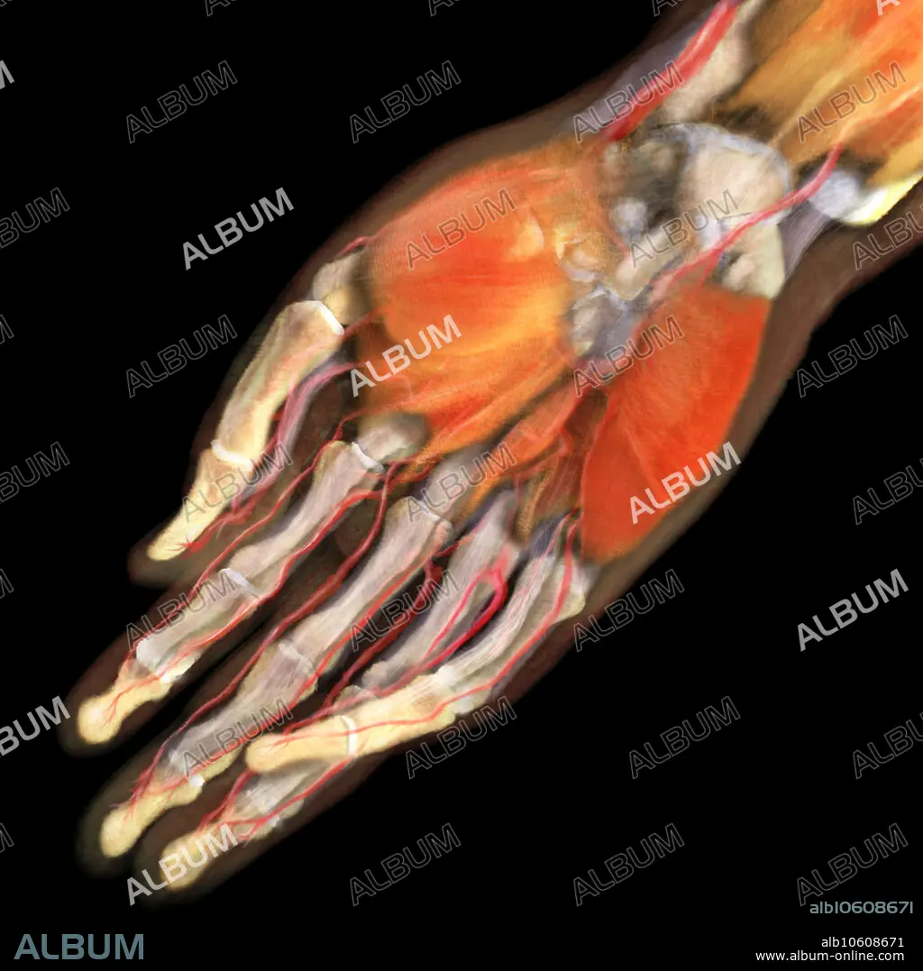
Right Hand Anatomy, Ventral View
Caption:
3D visualization based on scanned human data of the ventral side of a human right hand. On the palmar side, the thenar and hypothenar muscles are visible; lying beneath them are the metacarpal bones. The phalanges are revealed with the palmar digital arteries running also aside them.
Credit:
Album / Science Source / ANATOMICAL TRAVELOGUE
Releases:
Model: No - Property: No
Rights questions?
Rights questions?
Image size:
2000 x 2000 px | 11.4 MB
Print size:
16.9 x 16.9 cm | 6.7 x 6.7 in (300 dpi)
Keywords:
3D • ABDUCTOR • ANATOMICAL • ANATOMY • ANATOMY: BONES • ARTERIA • ARTERIAL • ARTERIE • ARTERIES • ARTERY • BIOLOGICAL • BIOLOGY • BONE • BREVIS • CARPAL • DIGITAL • DIGITI • DIMENSIONAL • DISTAL • EXTENSOR • EXTREMITY • FALANGE • FINGER • FINGERS • FLEXOR • GROSS ANATOMY • HAND • HEALTHY • HUMAN • HUMANE • HYPOTHENAR • INDIVIDUAL • INTERMEDIATE • LIMB • LUMBRICAL • METACARPAL • MINIMI • MUSCLE • MUSCULAR SYSTEM • MUSCULAR • MUSCULATURA • MUSCULATURE • MUSCULOSKELETAL • NORMAL • OF • OPPONENS • OSTEO-MUSCULAR • OSTEOMUSCULAR • PALM • PALMAR • PERSON • PHALANGE • PHALANGEAL • PHALANGES • PHALANX • PISIFORM • POLLICIS • RADIAL • RIGHT • SCAPHOID • SIDE • SKELETAL • STRONG • SYSTEM • TENDON • THE • THENAR • THREE • THREE-DIMENSIONAL • ULNAR • UPPER • VENTRAL • VIEW • VISUALIZATION • WRIST • ARTERIES
