alb3799303
Roentgen's X-Ray Machine, 19th Century
| Share |
|---|
 Pinterest Pinterest |
 Twitter Twitter |
 Facebook Facebook |
 Copy link Copy link |
 Email Email |
|
Add to another lightbox |
|
Add to another lightbox |


Buy this image.
Select the use:

Title: Roentgen's X-Ray Machine, 19th Century
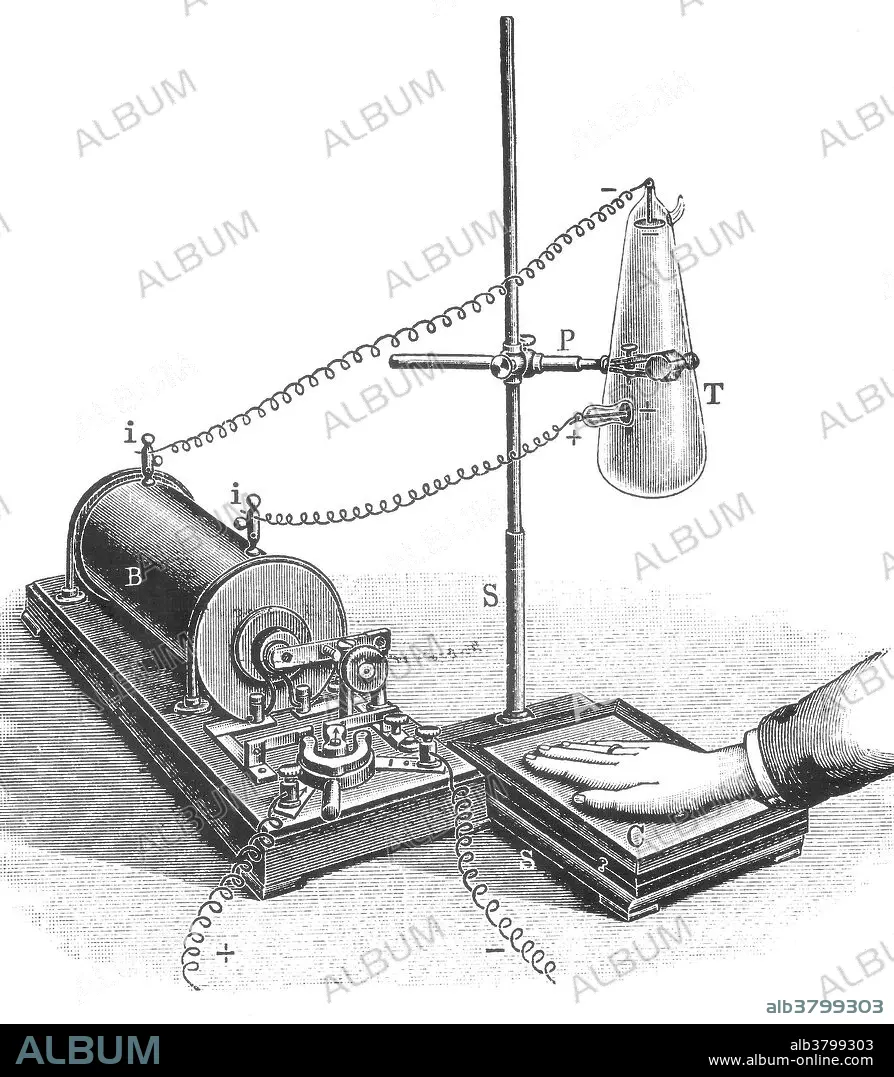
Caption: Wilhelm Conrad Roentgen (1845-1923), German experimental physicist and discoverer of X-rays. While using a discharge tube (in which an electric discharge is passed through a gas at low pressure) in a darkened room, Roentgen noticed that a card coated with barium platinocyanide glowed when the tube was switched on. The effect was not blocked by an intervening wall, or even a thin sheet of metal. Roentgen termed this newly discovered phenomenon X-ray radiation, and suggested that it consisted of electromagnetic rays with a shorter wavelength than light. He was awarded the first Nobel Prize for Physics in 1901. Drawing of the X-ray machine used by German physicist Wilhelm Roentgen to produce images of the hand. The generator (B) supplied a high voltage to the cathode ray tube (Crookes tube) at upper right (T). This tube produced X-rays which left an image of the hand on a covered, photographic plate (C).
Category: ILLUSTRATION • black & white • Medical: History
Credit: Album / Science Source / New York Public Library
Releases: ? Model Release: No - ? Property Release: No
Rights questions?
Rights questions?
Image size: 3082 × 3531 px | 31.1 MB
Print size: 26.1 × 29.9 cm | 1213.4 × 1390.2 in (300 dpi)
Keywords: 1845 • 1901 • 1923 • 19TH CENTURY • 20 20TH XX XXTH TWENTIETH CENTURY • 20 XX TWENTIETH CENTURY • 20TH CENTURY • 20TH • APPARATUS • ART • ARTWORK • BARIUM PLATINOCYANIDE • BARIUM • BLACK & WHITE • BW • CATHODE RAY TUBE • CELEBRITIES • CELEBRITY • CROOKES TUBE • DIAGNOSTIC IMAGING TECHNIQUE • DIAGNOSTIC TEST • DIAGNOSTIC • DISCOVERED • DRAWING • ELECTROMAGNETIC RADIATION • ELECTROMAGNETIC RAYS • ELECTROMAGNETIC • ENGRAVING • EQUIPMENT • EUROPEA • EUROPEAN • EUROPEANS • FAMOUS PEOPLE • FAMOUS • GERMAN • GERMANS • HISTORIC • HISTORICAL • HISTORY • ILLUSTRATION • ILLUSTRATIONS • ILUSTRATION • IMPORTANT • INSTRUMENT • MACHINE • MEDICAL IMAGING • MEDICAL PROCEDURE • MEDICAL • MEDICAL: HISTORY • MEDICINAL • NOBEL LAUREATE • NOBEL PRIZE LAUREATE • NOBEL PRIZE RECIPIENT • NOBEL PRIZE WINNER • NOBEL PRIZE • NOBEL RECIPIENT • NOBEL WINNER • NOBEL • NOBELIST • NON-INVASIVE TEST • NON-INVASIVE • NOTABLE • PHOTO PLATE • PHOTOGRAPHIC PLATE • PHYSICIST • PHYSICS • PLATINOCYANIDE • PROFESSOR • RADIOGRAM • RADIOGRAPHY • RADIOLOGY • ROENTGEN RAYS • ROENTGEN • RONTGEN RAYS • RONTGEN • SCIENCE • SHORT WAVE ELECTROMAGNETIC RAYS • SHORT WAVE • TOMOGRAM • TOMOGRAPHY • TOOL • TWENTIETH CENTURY • WAVELENGTH • WELL-KNOWN • WILHELM CONRAD ROENTGEN • WILHELM CONRAD RONTGEN • WILHELM KONRAD ROENTGEN • WILHELM KONRAD RONTGEN • WILHELM ROENTGEN • WILHELM RONTGEN • X RAY • X-RAY SCAN • X-RAY • XRAY SCAN • XRAY
