alb3886157
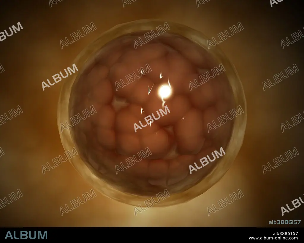
Microscopic view of a blastula during pregnancy.
| Share |
|---|
 Pinterest Pinterest |
 Twitter Twitter |
 Facebook Facebook |
 Copy link Copy link |
 Email Email |
|
Add to another lightbox |
|
Add to another lightbox |


Title:
Microscopic view of a blastula during pregnancy.
Caption:
Microscopic view of a blastula during pregnancy. After the cleavage has produced over 100 cells, the embryo is called a blastula. The blastula is usually a spherical layer of cells (the blastoderm) surrounding a fluid-filled or yolk-filled cavity (the blastocoel).
Credit:
Album / Stocktrek Images
Releases:
Model: No - Property: No
Rights questions?
Rights questions?
Image size:
4920 x 3690 px | 51.9 MB
Print size:
41.7 x 31.2 cm | 16.4 x 12.3 in (300 dpi)
Keywords:
ABSTRACT • ARTWORK • BALL • BIOMEDICAL ILLUSTRATIONS • BLASTOCOEL • BLASTOCYST • BLASTODERM • BLASTULA • CELL BIOLOGY • CELL POLARITY • CELL • CIRCLE • CLOSE UP • CLOSE-UP • CLOSEUP • COLOR IMAGE • COLOUR IMAGE • CONCEPTUS • CYTOLOGY • DETAIL • DEVELOPMENT • DIGITALLY GENERATED IMAGE • EGG YOKE • EMBRYO • EMBRYOBLAST • EMBRYOGENESIS • ENLARGEMENT • FETUS • FLUID • FOETUS • FOREGROUND • GENE EXPRESSION • HEALTHCARE • HOLLOW • HORIZONTAL • ILLUSTRATION • ILLUSTRATIONS • INNER CELL MASS • MAGNIFICATION • MEDICAL • MEDICINAL • MEDICINE • MICROBIOLOGY • MICROSCOPIC • MOLECULAR BIOLOGY • MÓRULA • NO PEOPLE • PLURIBLAST • PREGANCY • PREGNANCY • ROUND • SCIENCE • SINGLE OBJECT • SPHÈRE • THREE DIMENSIONAL • TRANSLUCENT • TRANSPARENT • WITHOUT PEOPLE • YOLK
