alb10655682
Normal neck and cervical spine, MRI
| Share |
|---|
 Pinterest Pinterest |
 Twitter Twitter |
 Facebook Facebook |
 Copy link Copy link |
 Email Email |
|
Add to another lightbox |
|
Add to another lightbox |


Title:
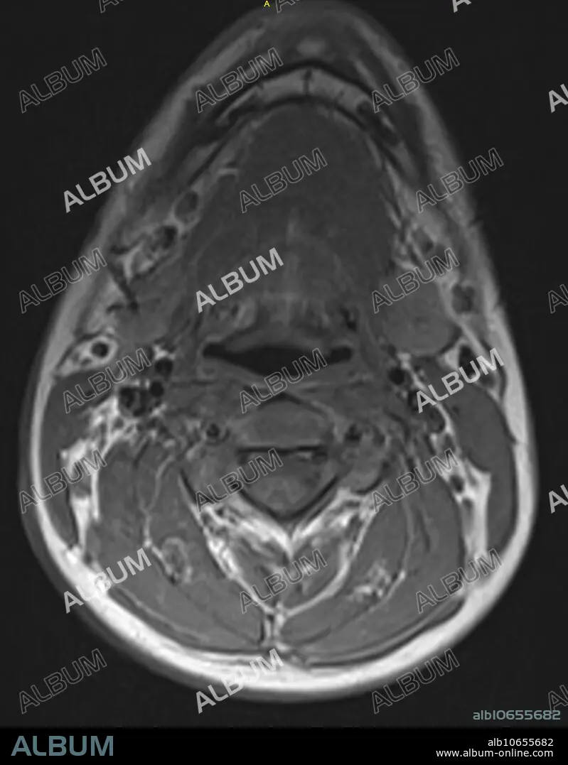
Normal neck and cervical spine, MRI
Caption:
Normal T1 weighted axial MRI showing the neck and cervical spine in a 16 year old male at the level of C3.
Credit:
Album / Science Source / Steven Needell
Releases:
Model: No - Property: No
Rights questions?
Rights questions?
Image size:
1716 x 2200 px | 10.8 MB
Print size:
14.5 x 18.6 cm | 5.7 x 7.3 in (300 dpi)
Keywords:
ABNORMAL • ANATOMICAL • ANATOMY • ANATOMY: BONES • ARTERIA • ARTERY • AXIAL • AXIS • BODY • BONE • C-SPINE • CANAL • CAP • CERVICAL • CONDITION • CORD • CROSS • CROSS-SECTIONAL • CT • DIAGNOSTIC • DISEASE • DISORDER • FAT • FLUID • GROSS ANATOMY • IMAGE • IMAGING • L-SPINE • LSP • LUMBAR • LUMBORUM • MAGNETIC • MEDICAL • MEDICINAL • MEDICINE • MRI • NECK • NECKS • NERVE • NORMAL • ORGAN • PATHOLOGICAL • PATHOLOGY • RADIOLOGY • RESONANCE • SCAN • SECTION • SECTIONAL • SHORT • SOFT • SONORITY • SPINAL • SPINE • T-SPINE • THORACIC • THROAT • TISSUE • TRANSVERSE • TSP • UNHEALTHY • UPPER • VEIN • VENA
