alb10660588
Tongue cancer, PET CT scan
| Share |
|---|
 Pinterest Pinterest |
 Twitter Twitter |
 Facebook Facebook |
 Copy link Copy link |
 Email Email |
|
Add to another lightbox |
|
Add to another lightbox |


Buy this image.
Select the use:

Title:
Tongue cancer, PET CT scan
Caption:
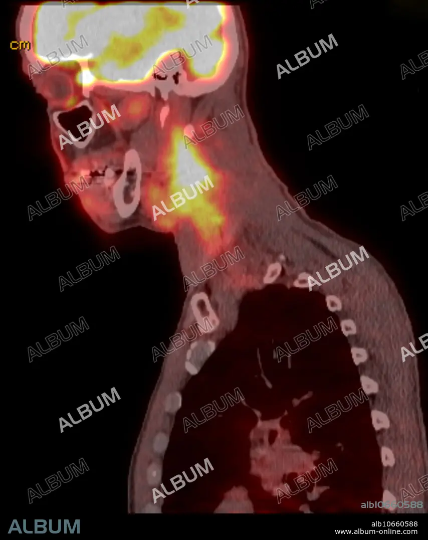
Positron emission tomography of 60 yo male with right squamous cell carcinoma of the tongue. Sagittal FDG CT PET scan reveals large conglomeration of lymph nodes within the right neck beginning at the angle of the mandible and extending to the level lower neck (level 5) and also extension into the posterior triangle. The mass is deep to the sternocleidomastoid. Mass extends posteriorly into the posterior triangle. Corresponding intense metabolic uptake with SUV of 6.0. There is focal asymmetric uptake involving the base the tongue, with SUV of 4.9.
Personalities:
Credit:
Album / Science Source / Steven Needell
Releases:
Image size:
1454 x 1744 px | 7.3 MB
Print size:
12.3 x 14.8 cm | 4.8 x 5.8 in (300 dpi)
Keywords:
60 • ABNORMAL • ANIMAL: CAT • ARTERIA • ARTERY • BLEEDING • CANCER • CARCINOMA • CAROTID • CAT • CELL • COMPUTED • CONDITION • CONFLUENT • CROSS-COUNTRY VEHICLE • CT • DIAGNOSTIC • DISEASE • DISORDER • ÉMISSION • FELIS CATUS • FOUR-WHEEL DRIVE • HAEMORRHAGE • HEMMORHAGE • HEMORRHAGE • HEMORRHAGED • HEMORRHAGING • HYPOPHARYNX • IMAGING • INFILTRATING • LYMPHADENOPATHY • LYMPHOMA • MALE • MANDIBLE • MARGINATED • MASS • MEDICAL • MEDICINAL • MEDICINE • MESS • MESSY • METASTATIC • MPR • MUSCLE • NASOPHARYNGEAL • NECK • NECKS • NECROSIS • PATHOLOGICAL • PATHOLOGY • PET • POSITRON • RADIOLOGY • SAGGITAL • SAGITTAL VIEW • SAGITTAL • SCAN • SOFT • SPREAD • SQUAMOUS • STERNOCLEIDO-MASTOID • STERNOCLEIDOMASTOID MUSCLE • STERNOCLEIDOMASTOID • SUV • THROAT • THYROID • TISSUE • TOMOGRAPHY • TONGUE • TUMOR • TUMOUR • UNHEALTHY • VOLUME
