alb3783901
X-ray of large intestine
| Share |
|---|
 Pinterest Pinterest |
 Twitter Twitter |
 Facebook Facebook |
 Copy link Copy link |
 Email Email |
|
Add to another lightbox |
|
Add to another lightbox |


Buy this image.
Select the use:

Title:
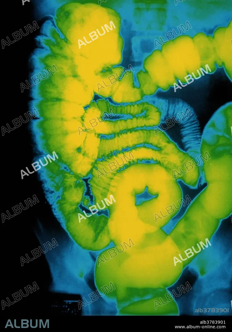
X-ray of large intestine
Caption:
Large intestine. Colored X-ray image of the human abdomen showing the large intestine (colon), after a barium enema. At center are loops of the small intestine. These lead into the dilated colon, of which there are three regions. At left is the ascending region of the colon; at top, the transverse colon runs horizontally, leading to the descending colon (at right). At bottom center is the rectum. The colon absorbs water and electrolytes from digested food; periodic muscular contractions move the dehydrated contents (feces) towards the rectum. A barium enema is a suspension of barium sulphate (which is opaque to X-rays) that is infused into the rectum.
Credit:
Album / Science Source / Susan Leavines
Releases:
Model: No - Property: No
Rights questions?
Rights questions?
Image size:
3168 x 4308 px | 39.0 MB
Print size:
26.8 x 36.5 cm | 10.6 x 14.4 in (300 dpi)
Keywords:
ABDOMEN • ANATOMY • BARIUM ENEMA • BARIUM X-RAY • BARIUM X-RAYS • BELLY • BOWELS • COLON • COLONS • DIGESTIVE SYSTEM • GREEN • GROSS ANATOMY • GUT • HEALTH • HUMAN ABDOMEN • HUMAN BODIES • HUMAN BODY • INTESTINE • INTESTINES • LARGE INTESTINE • LARGE INTESTINES • MEDICAL • MEDICINAL • MEDICINE • PHYSIOLOGIE • PHYSIOLOGY • SMALL INTESTINE • SMALL INTESTINES • STOMACH • VERTICAL LINES • VERTICAL • X-RAY • X-RAYS
