alb3798168
Mitosis, Prophase, LM
| Share |
|---|
 Pinterest Pinterest |
 Twitter Twitter |
 Facebook Facebook |
 Copy link Copy link |
 Email Email |
|
Add to another lightbox |
|
Add to another lightbox |


Title:
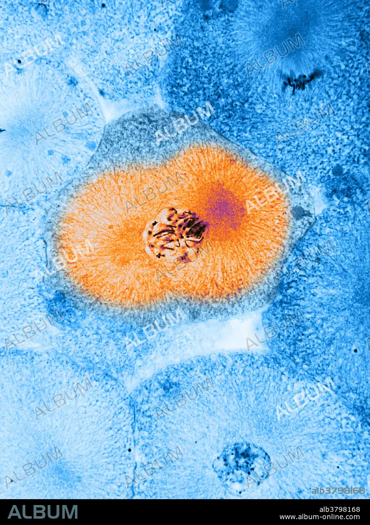
Mitosis, Prophase, LM
Caption:
Colour enhanced light micrograph showing mitosis - prophase, in whitefish blastula, the centriole has divided and the daughter centrioles have moved to opposite poles of the cell, a centriole plus the spindle fibers. Magnification 1,100x. Mitosis, the usual method of cell division, characterized typically by the resolving of the chromatin of the nucleus into a threadlike form, which condenses into chromosomes, each of which separates longitudinally into two parts, one part of each chromosome being retained in each of two new cells resulting from the original cell. The four main phases of mitosis are prophase, metaphase, anaphase, and telophase. Blastula, an animal embryo at the stage immediately following the division of the fertilized egg cell, consisting of a ball-shaped layer of cells around a fluid-filled cavity known as a blastocoel. A centriole is a small, cylindrical cell organelle, seen near the nucleus in the cytoplasm of most eukaryotic
Credit:
Album / Science Source / BIOPHOTO ASSOCIATES
Releases:
Model: No - Property: No
Rights questions?
Rights questions?
Image size:
3601 x 4851 px | 50.0 MB
Print size:
30.5 x 41.1 cm | 12.0 x 16.2 in (300 dpi)
Keywords:
ANATOMY • ANIMAL • BIOLOGY • BLASTULA • BREEDING • CELL • CELLULAR • CENTRIOLE • COLORIZED • CONCEPTUS • CYTOLOGY • CYTOPLASM • DIVISION • EMBRYO • ENHANCED • EUKARYOTE • EUKARYOTIC • FETUS • FIBERS • FOETUS • GROSS ANATOMY • HISTOLOGY • LIGHT • LM • MICROGRAPH • MICROGRAPHY • MICROSCOPIC • MICROSCOPY • MITOSIS • NO ONE • NO-ONE • NOBODY • ORGANELLE • PHYSIOLOGIE • PHYSIOLOGY • PROMETAPHASE • PROPHASE • REPRODUCTION • SPINDLE • STRUCTURE • WHITEFISH
