alb3784676
Anatomy of Human Eye
| Share |
|---|
 Pinterest Pinterest |
 Twitter Twitter |
 Facebook Facebook |
 Copy link Copy link |
 Email Email |
|
Add to another lightbox |
|
Add to another lightbox |


Buy this image.
Select the use:

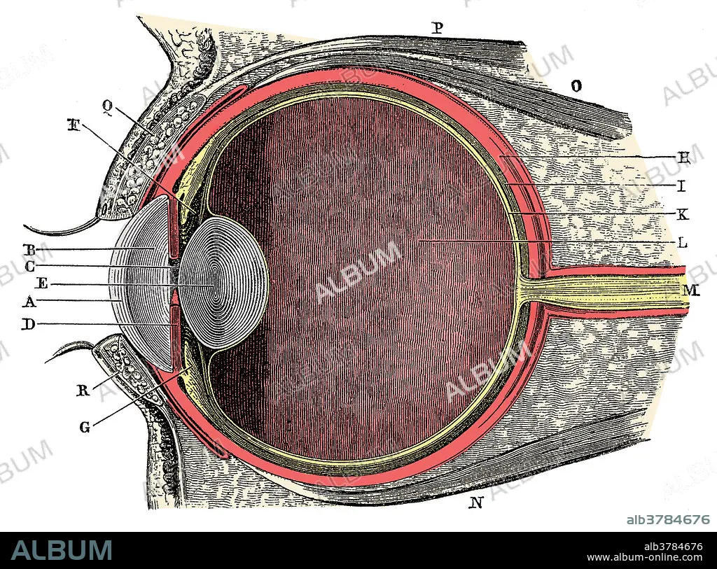
Title: Anatomy of Human Eye
Caption: Anatomy of the Human Eye. Cornea (A), aqueous humor (B), iris (C), pupil (D), lens (E), Suspensory ligament (F), ciliary body (G), sclera (H), choroid (I), retina (K), vitreous humor (L), optic nerve (M). This is a historical anatomical illustration from the 1890's.
Credit: Album / SCIENCE SOURCE
Releases: ? Model Release: No - ? Property Release: No
Rights questions?
Rights questions?
Image size: 3717 × 2760 px | 29.4 MB
Print size: 31.5 × 23.4 cm | 1463.4 × 1086.6 in (300 dpi)
Keywords: ANATOMICAL • ANATOMY • AQUEOUS • ART • ARTWORK • BODY • CHOROID • CILIARY • COLORED • CORNEA • DRAWING • ENHANCED • EYE • EYEBALLS • EYES • GROSS ANATOMY • HISTORICAL • HUMAN • HUMANE • HUMOR • HUMOUR • ILLUSTRATION • ILLUSTRATIONS • ILUSTRATION • INDIVIDUAL • IRIS • LENS • LIGAMENT • LIGAMENTUM • MEDICAL • MEDICINAL • NERVE • OPTIC • PERSON • PUPIL • RETINA • RETINAE • RETINAS • SCLERA • SUSPENSORY • VITREOUS
