alb3788974
Top View of Normal Brain, Illustration
| Share |
|---|
 Pinterest Pinterest |
 Twitter Twitter |
 Facebook Facebook |
 Copy link Copy link |
 Email Email |
|
Add to another lightbox |
|
Add to another lightbox |


Buy this image.
Select the use:

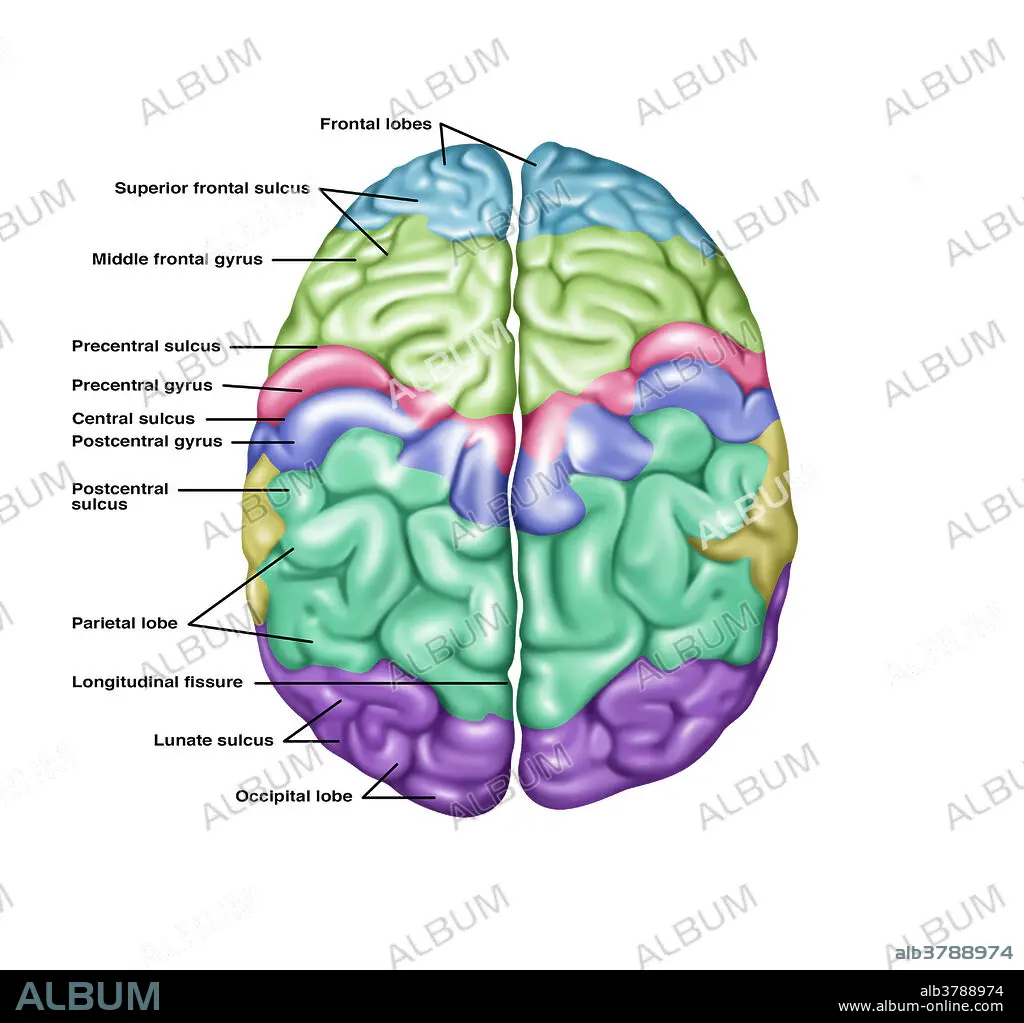
Title: Top View of Normal Brain, Illustration
Caption: Illustration showing anatomy of a normal brain in a superior (top) view. Noted from top to bottom on the left side are the following: frontal lobes, superior frontal sulcus, middle frontal gyrus, precentral sulcus, precentral gyrus, central sulcus, postcentral gyrus, postcentral sulcus, parietal lobe, longitudinal fissure, lunate sulcus, and the occipital lobe.
Credit: Album / Science Source / Gwen Shockey
Releases: ? Model Release: No - ? Property Release: No
Rights questions?
Rights questions?
Image size: 4337 × 4110 px | 51.0 MB
Print size: 36.7 × 34.8 cm | 1707.5 × 1618.1 in (300 dpi)
Keywords: ANATOMY • ANNOTATED • ARTWORK • BRAIN • CENTRAL • DRAWING • FISSURE • FRONT • FRONTAL • GRAPHIC • GROSS ANATOMY • GYRUS • HEALTHY • ILLUSTRATION • ILLUSTRATIONS • ILUSTRATION • INFOGRAPHIC • INFORMATION • INTERHEMISPHERIC • LOBE • LOBULE • LONGITUDINAL • LUNATE • MIDDLE • NORMAL • OCCIPITAL • OCCIPITALIS • PARIETAL • POSTCENTRAL • PRECENTRAL • SULCUS • SUPERIOR • VIEW
