alb10654268
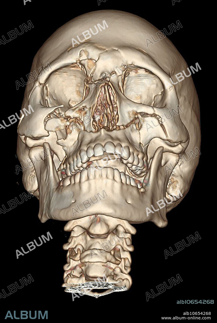
3D CT of Lefort Facial Fractures

|
Add to another lightbox |
|
Add to another lightbox |


Title:
3D CT of Lefort Facial Fractures
Caption:
This frontal view from a 3D CT reconstruction shows extensive facial fractures representing bilateral Lefort II fractures and right sided (on viewers left) Lefort III fractures.
Credit:
Album / Living Art Enterprises, LLC/Science Source
Releases:
Model: No - Property: No
Rights questions?
Rights questions?
Image size:
4200 x 5940 px | 71.4 MB
Print size:
35.6 x 50.3 cm | 14.0 x 19.8 in (300 dpi)
Keywords:
3D • ANATOMY: BONES • ANATOMY: SKULL • ANIMAL: CAT • AXIAL • BONE • BONES • CAT • COMPUTED • CRANEO • CRANEOS • CRANIUM • CRANIUMS • CT • FACE • FACIAL • FELIS CATUS • FRACTURE • FRACTURES • HIT • II • III • IN • INJURY • LEFORT • MEDICAL • MEDICINAL • OF • REMAINS (SKELETON) • SCAN • SÉVÈRE • SKELETON • SKULL • SKULL, ANATOMY • SKULLS • TO • TOMOGRAPHY • TRAUMA
 Pinterest
 Twitter
 Facebook
 Copy link
 Email
Email

