alb3783541
Heart, External Anatomy, Illustration
| Share |
|---|
 Pinterest Pinterest |
 Twitter Twitter |
 Facebook Facebook |
 Copy link Copy link |
 Email Email |
|
Add to another lightbox |
|
Add to another lightbox |


Buy this image.
Select the use:

Title: Heart, External Anatomy, Illustration
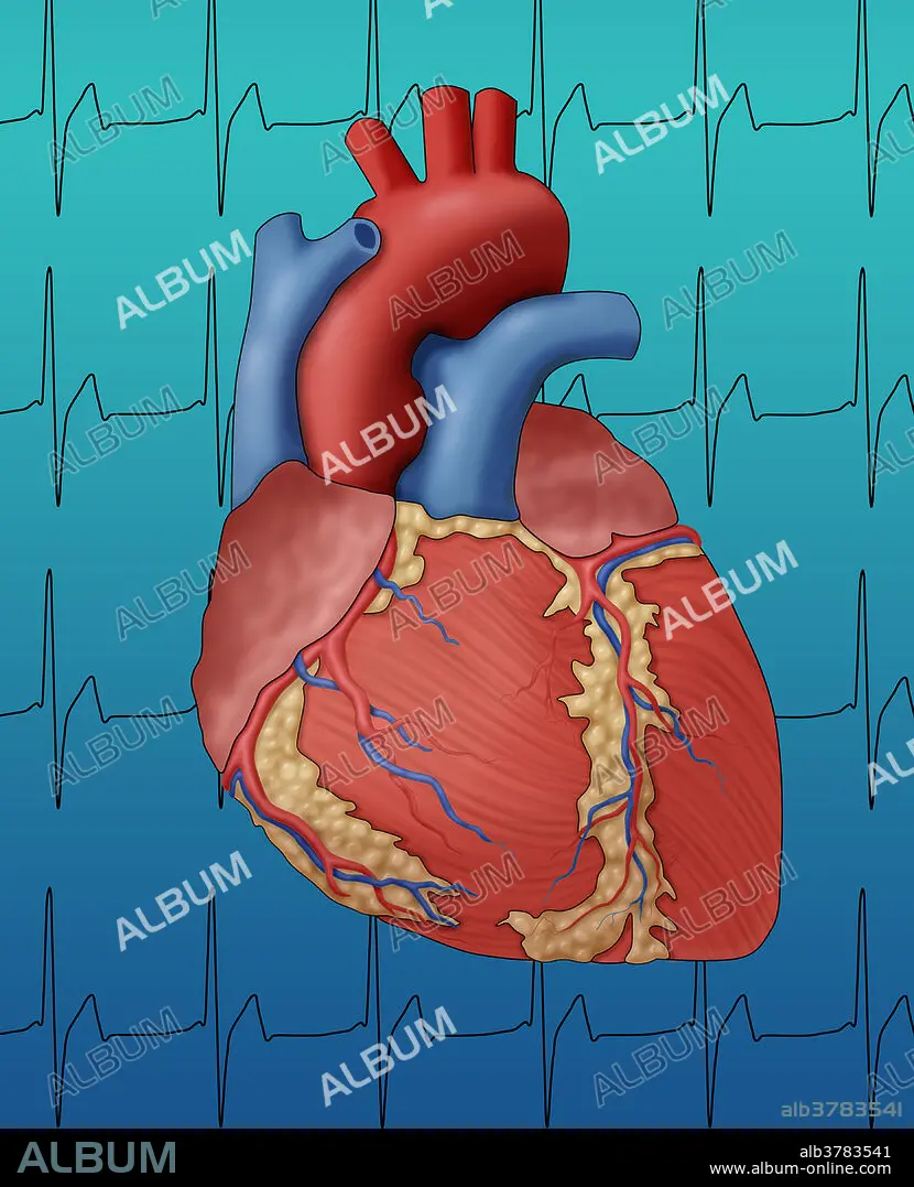
Caption: Illustration showing the external anatomy of a human heart, seen from the front. The surface blood vessels are the coronary arteries (bright red) and veins (blue), bringing oxygenated blood to the heart and removing deoxygenated blood. This healthy heart has a normal heart rate, depicted by an EKG is in the background.
Credit: Album / Science Source / Monica Schroeder
Releases: ? Model Release: No - ? Property Release: No
Rights questions?
Rights questions?
Image size: 3732 × 4604 px | 49.2 MB
Print size: 31.6 × 39.0 cm | 1469.3 × 1812.6 in (300 dpi)
Keywords: ANATOMICAL • ANATOMY • ANTERIOR • ANTERIORLY • AORTA • ART • ARTERIA • ARTERIAL • ARTERIE • ARTERIES • ARTERY • ARTWORK • BIOLOGY • BLOOD • CARDIAC • CARDIOLOGY • CARDIOVASCULAR • CAROTID • CAVA • CAVAE • CONCEPT • CONCEPTUAL • CORONARY • ECG • EKG • ELECTROCARDIOGRAM • EXTERNAL • FRONT • FRONTAL • GROSS ANATOMY • HEALTHY • HEART • ILLUSTRATION • ILLUSTRATIONS • ILUSTRATION • MEDICAL • MEDICINAL • MUSCLE • NORMAL • OF • ORGAN • PULMONARY • RATE • SANGUINE • VEIN • VEINS • VENA • VENTRICLES • VESSEL • VESSELS • ARTERIES
