alb10671850
Intracranial Dermoid Cyst on MRI
| Share |
|---|
 Pinterest Pinterest |
 Twitter Twitter |
 Facebook Facebook |
 Copy link Copy link |
 Email Email |
|
Add to another lightbox |
|
Add to another lightbox |


Buy this image.
Select the use:

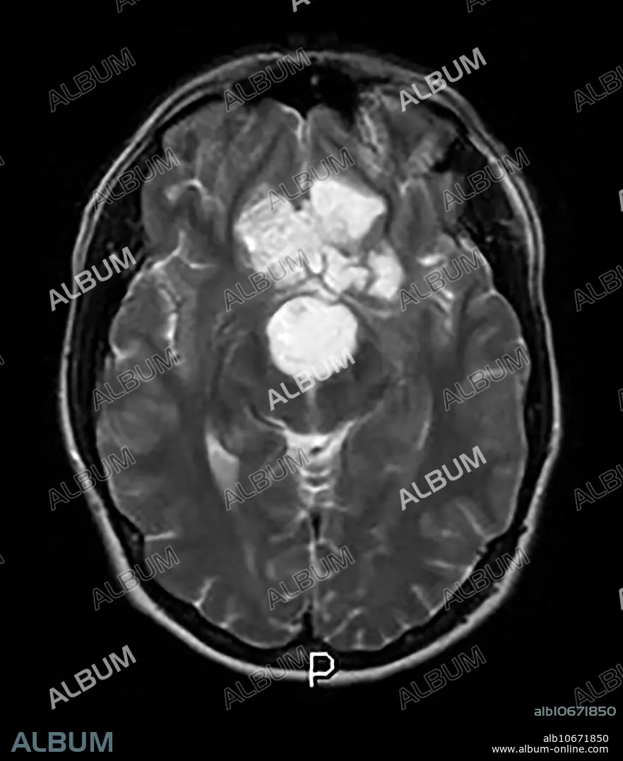
Title: Intracranial Dermoid Cyst on MRI
Caption: This axial (cross sectional) T2 weighted MR image shows a mass lesion with heterogeneous signal in the inferior frontal region which represents a benign, congenital Dermoid Cyst.
Personalities: MASS
Credit: Album / Living Art Enterprises, LLC/Science Source
Releases: ? Model Release: No - ? Property Release: No
Rights questions?
Rights questions?
Image size: 3900 × 4530 px | 50.5 MB
Print size: 33.0 × 38.4 cm | 1535.4 × 1783.5 in (300 dpi)
Keywords: ABNORMAL • BRAIN • CONGENITAL • CYST • DERMOID • EPIDERMOID • FAT • FATTY • INTRACRANIAL • MASS • MRI • TUMOUR • WITH
