alb3808882
Gangrene, Illustration, 1830s
| Share |
|---|
 Pinterest Pinterest |
 Twitter Twitter |
 Facebook Facebook |
 Copy link Copy link |
 Email Email |
|
Add to another lightbox |
|
Add to another lightbox |


Buy this image.
Select the use:

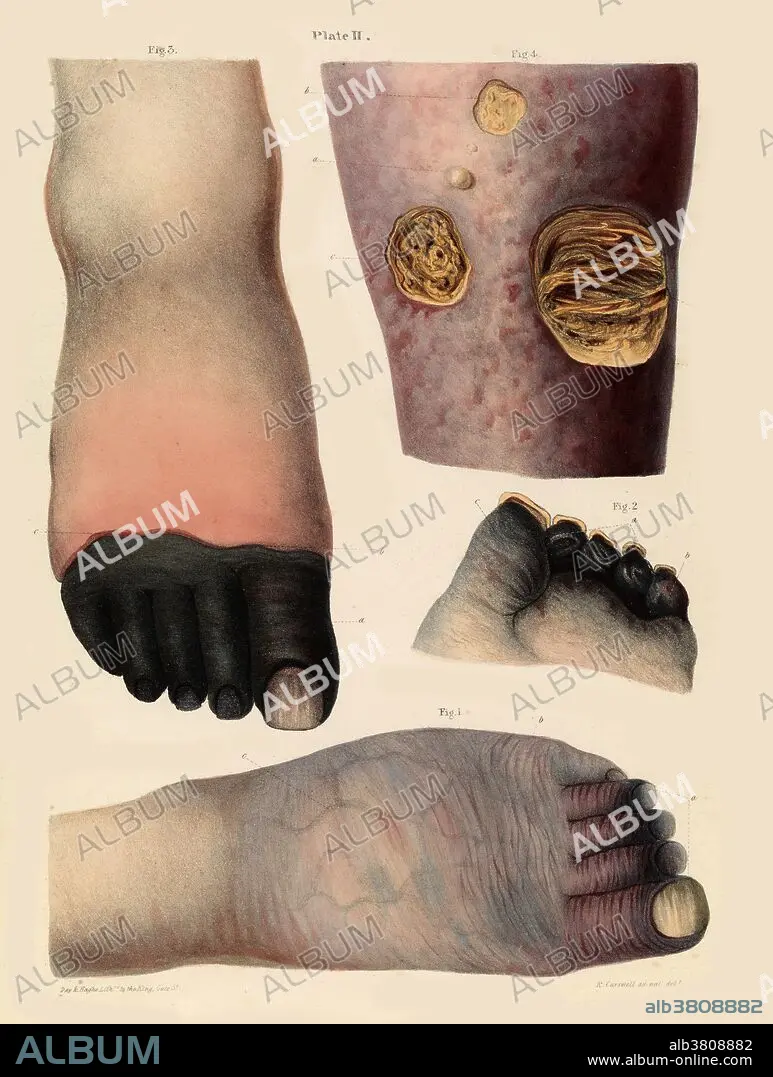
Title: Gangrene, Illustration, 1830s
Caption: Gangrene illustration from the 1830s. Figs 1 and 2 (bottom and centre right) show gangrena senilis - discoloration of the toes at the beginning of the disease. Fig 3 (top left) shows gangrene of the toes. Fig 4 (top right) shows mortification of the skin and subjacent cellular tissue from an obstacle to the return of the venous blood in consequence of disease of the heart. Robert Carswell.
Category: ILLUSTRATION • Medical: History
Credit: Album / Science Source / Wellcome Images
Releases: ? Model Release: No - ? Property Release: No
Rights questions?
Rights questions?
Image size: 4952 × 6556 px | 92.9 MB
Print size: 41.9 × 55.5 cm | 1949.6 × 2581.1 in (300 dpi)
Keywords: 1800S • 19TH CENTURY • ABNORMAL • ADVANCED • ANATOMY: LEG • ART • ARTWORK • BLACK GANGRENE • CONDITION • DERMATOLOGICAL • DERMATOLOGY • DISCOLORATION • DISEASE • DISORDER • DRAWING • DRY GANGRENE • FOOT FEET • FOOT • GANGRENA SENILIS • GANGRENE • GANGRENOUS NECROSIS • HISTORIC • HISTORICAL • HISTORY • ILLUSTRATION • ILLUSTRATIONS • ILUSTRATION • LEG • LEG, ANATOMY • LEGS • MEDICAL • MEDICAL: HISTORY • MEDICINAL • MEDICINE • MESS • MESSY • MOIST GANGRENE • MORTIFICATION • NECROSIS • PATHOLOGY • PATIENT • PEOPLE • PERSON • SÉVÈRE • SKIN DISEASE • SKIN • TOE • TOES • WET GANGRENE
