alb9202346
Illustration of Skin Section
| Share |
|---|
 Pinterest Pinterest |
 Twitter Twitter |
 Facebook Facebook |
 Copy link Copy link |
 Email Email |
|
Add to another lightbox |
|
Add to another lightbox |


Buy this image.
Select the use:

Title: Illustration of Skin Section
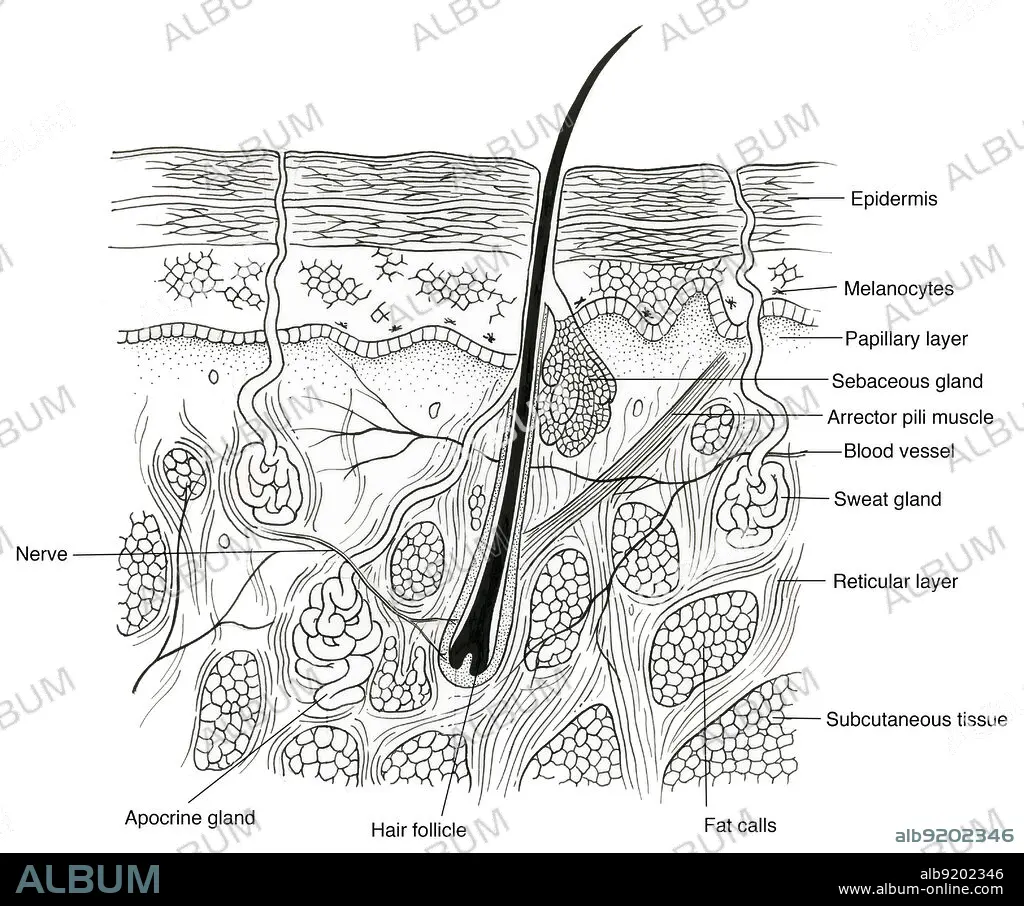
Caption: Anatomical illustration of a section through human skin, showing the epidermis, melanocytes, papillary layer, sweat gland, hair follicle, blood vessels, fat cells, sebaceous gland, arrector pili muscle, reticular layer, apocrine gland, subcutaneous tissue, and nerve.
Credit: Album / Science Source
Releases: ? Model Release: No - ? Property Release: No
Rights questions?
Rights questions?
Image size: 4332 × 3609 px | 44.7 MB
Print size: 36.7 × 30.6 cm | 1705.5 × 1420.9 in (300 dpi)
Keywords: ANATOMY • APOCRINE • ARRECTOR • ART • BLOOD • BODY • BT2994 • BW • CELL • CROSS CUT • CROSS-CUT • CROSS-SECTION • DERMIS • EPIDERMIS • FAT • FATTY • FOLLICLE • GLAND • GROSS ANATOMY • HAIR • HEALTHY • HUMAN • HUMANE • ILLUSTRATION • ILLUSTRATIONS • ILUSTRATION • INDIVIDUAL • LAYER • MELANOCYTES • MUSCLE • NERVE • NERVOUS • NORMAL • PAPILLARY • PERSON • PILI • RETICULAR • RF • SANGUINE • SCARFSKIN • SEBACEOUS • SECTION • SECTIONAL • SKIN • STRUCTURE • SUBCUTANEOUS • SWEAT • SYSTEM • TISSUE • VESSEL
