alb3777959
Head and Brain Anatomy
| Share |
|---|
 Pinterest Pinterest |
 Twitter Twitter |
 Facebook Facebook |
 Copy link Copy link |
 Email Email |
|
Add to another lightbox |
|
Add to another lightbox |


Buy this image.
Select the use:

Title:
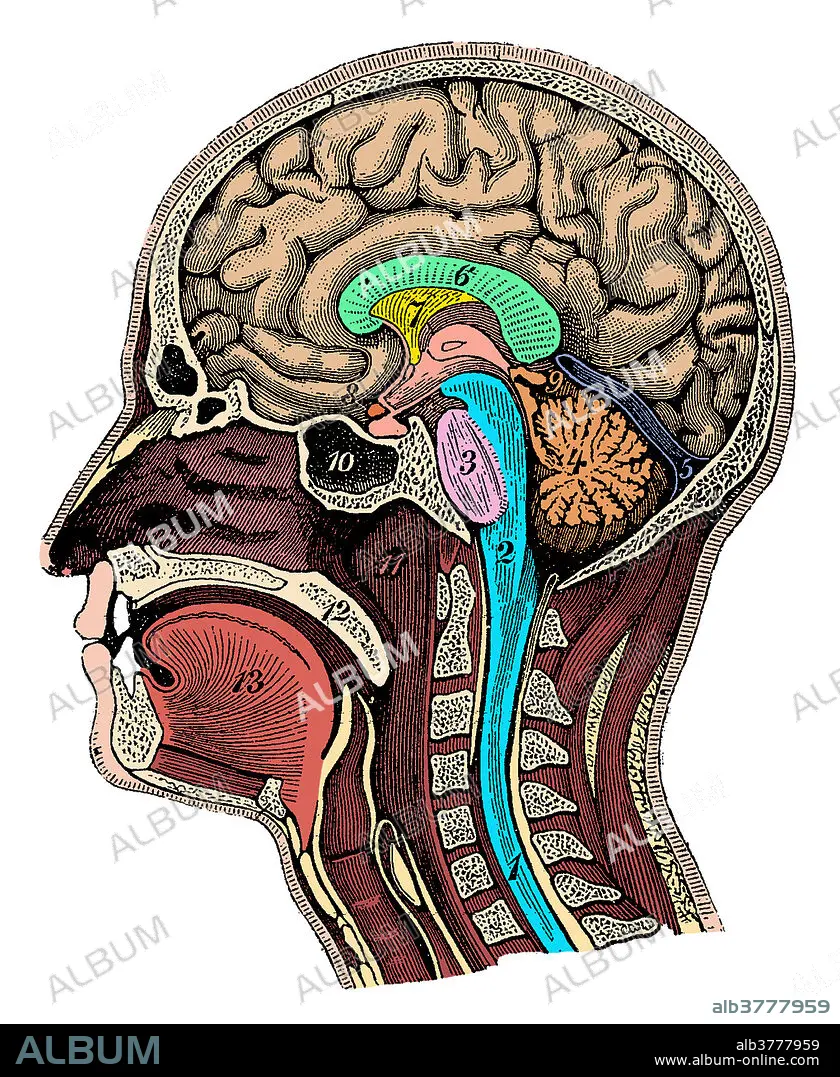
Head and Brain Anatomy
Caption:
Color enhanced illustration of a cross-section of the head showing the brain showing the spinal cord, medulla oblongata, pons, cerebellum, transverse fissure, corpus callosum, septum pellucidum, optic chiasm, pineal gland, sphenoid sinus, nasopharynx, soft palate and tongue. This is an historical illustration from the 1890's.
Credit:
Album / SCIENCE SOURCE
Releases:
Image size:
2850 x 3475 px | 28.3 MB
Print size:
24.1 x 29.4 cm | 9.5 x 11.6 in (300 dpi)
Keywords:
ANATOMICAL • ANATOMY • BRAIN • BULB • CALLOSUM • CENTRAL • CEREBELLUM • CHIASM • CORD • CORPUS • CROSS-SECTIONAL • FISSURE • GLAND • GROSS ANATOMY • HEAD • HISTORICAL • HUMAN • HUMANE • ILLUSTRATION • ILLUSTRATIONS • INDIVIDUAL • MEDICAL • MEDICINAL • MEDULLA • NASOPHARYNX • NERVOUS • OBLONGATA • OPTIC • PALATE • PELLUCIDUM • PERSON • PINEAL • PITH • PONS • SEPTUM • SINUS • SOFT • SPHENOID • SPINAL • SYSTEM • TONGUE • TRANSVERSE
