alb10666019
Tilia stem, LM
| Share |
|---|
 Pinterest Pinterest |
 Twitter Twitter |
 Facebook Facebook |
 Copy link Copy link |
 Email Email |
|
Add to another lightbox |
|
Add to another lightbox |


Buy this image.
Select the use:

Title:
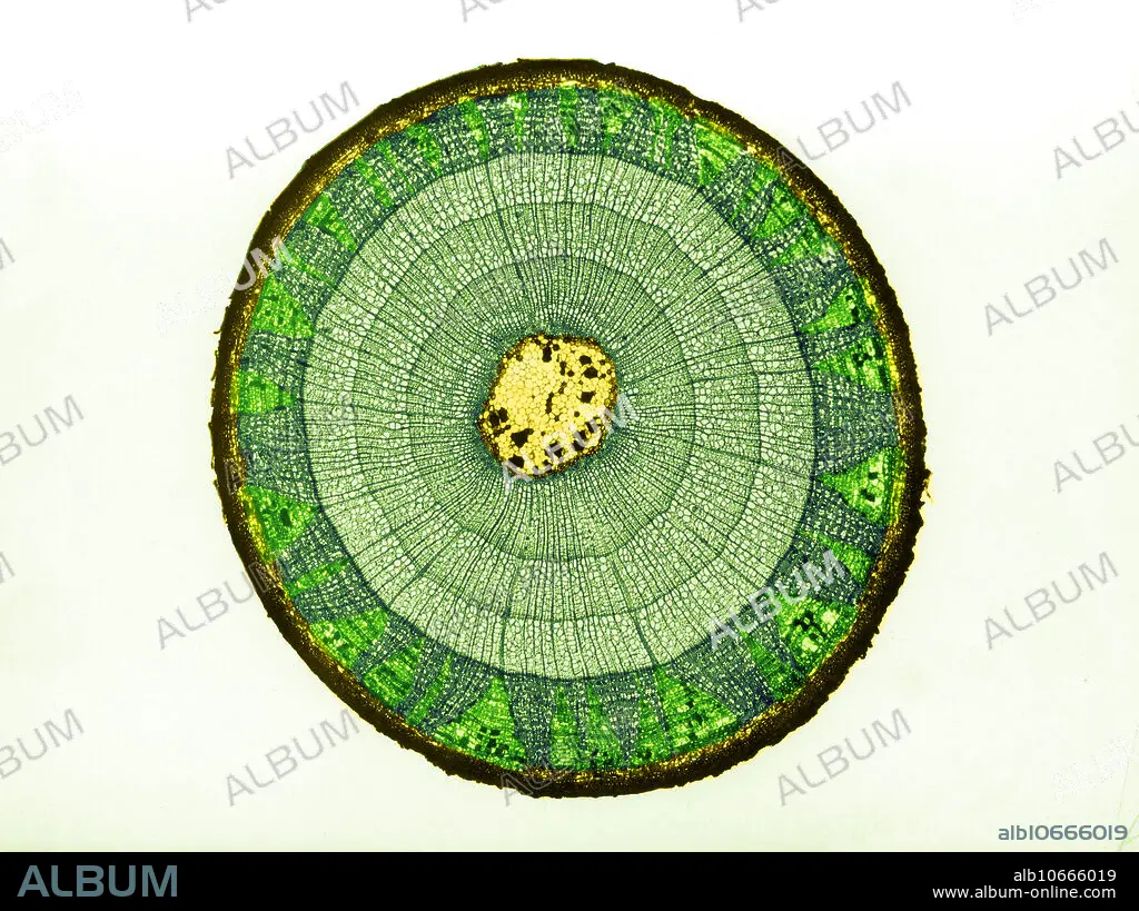
Tilia stem, LM
Caption:
Colour enhanced light micrograph of the cross section of a Tilia sp. stem. The stem is three-years-old as it has three growth rings (concentric circles). The following is depicted (from outer to inner circle): epidermis (dark yellow edge), primary and secondary phloem (dark green section) with pith rays (light green triangular parts in phloem), primary and secondary Xylem (lighter sets of rings), and Pith (yellow portion in centre).
Credit:
Album / Science Source / Omikron
Releases:
Model: No - Property: No
Rights questions?
Rights questions?
Image size:
4217 x 3154 px | 38.1 MB
Print size:
35.7 x 26.7 cm | 14.1 x 10.5 in (300 dpi)
Keywords:
ANATOMY • ANGIOSPERM • BIOLOGY • BOTANICAL • BOTANY • BOTANY • BOTANY • CAMBIUM • CELL • CELLULAR • COLORIZED • CORK • CORTEX • ENHANCED • ENHANCEMENT • EUDICOT • FIBER • FIBERS • FLORA • GROSS ANATOMY • GROWTH • INNER • LIGHT • LM • MEDULLA • MICROBIOLOGY • MICROGRAPH • MICROGRAPHY • NO ONE • NO-ONE • NOBODY • PARENCHYMA • PHLOEM • PITH • PLANT • PLATE • PLATES • PRIMARY • RING • RINGS • SCIENCE • SIEVE • STEM • STRUCTURE • TILIA • TISSUE • VESSEL • XYLEM
