alb9201983
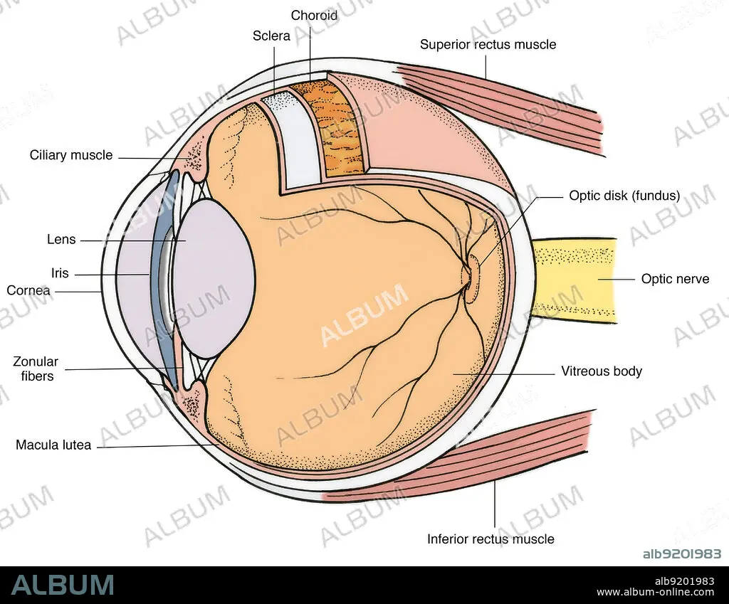
Illustration of Eye Anatomy
| Share |
|---|
 Pinterest Pinterest |
 Twitter Twitter |
 Facebook Facebook |
 Copy link Copy link |
 Email Email |
|
Add to another lightbox |
|
Add to another lightbox |


Buy this image.
Select the use:

Title: Illustration of Eye Anatomy
Caption: Anatomical illustration of eye and eyeball (lateral view), showing (on eye) pupil, iris, scerla, and caruncula, and (on eyeball) ciliary muscle, lens, iris, pupil, zonular fibers, macula lutea, inferior rectus muscle, vitreous body, optic nerve, optic disk (fundus), superior rectus muscle, choroid, sclera, and ciliary muscle.
Credit: Album / Science Source / Science Source
Releases: ? Model Release: No - ? Property Release: No
Rights questions?
Rights questions?
Image size: 4548 × 3534 px | 46.0 MB
Print size: 38.5 × 29.9 cm | 1790.6 × 1391.3 in (300 dpi)
Keywords: ANATOMY • ART • BODY • BT7961 • CARUNCULA • CHOROID • CILIARY • COLOR • COLORIZED • DISC • DISCUS • DISK • ENHANCE • ENHANCED • EPROMO-375 • EYE • EYEBALL • EYEBALLS • EYES • FIBERS • FUNDI • FUNDUS • GROSS ANATOMY • HEALTHY • HUMAN • HUMANE • ILLUSTRATION • ILLUSTRATIONS • ILUSTRATION • INDIVIDUAL • INFERIOR • IRIS • LENS • LUTEA • MACULA • MEDICAL • MEDICINAL • MUSCLE • NERVE • NORMAL • OPTIC • PERSON • PUPIL • RECTUS • RF • SCERLA • SCLERA • STRUCTURE • SUPERIOR • VITREOUS • ZONULAR
