alb3772973
Lungs and Pleura, illustration
| Share |
|---|
 Pinterest Pinterest |
 Twitter Twitter |
 Facebook Facebook |
 Copy link Copy link |
 Email Email |
|
Add to another lightbox |
|
Add to another lightbox |


Buy this image.
Select the use:

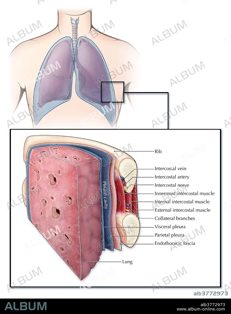
Title: Lungs and Pleura, illustration
Caption: An illustrated section of a lung and the ribcage showcasing different layers of tissue and muscle. The pleura (blue) is a serous membrane covering the lungs (visceral pleura) and chest wall (parietal pleura), creating a fluid filled space (pleural cavity) which lubricates the lungs to aid in breathing. Between each rib are a series of intercostal muscles, arteries, veins and nerves. A layer of endothoracic fascia also lines the inner surface of the ribcage.
Credit: Album / Science Source / Evan Oto
Releases: ? Model Release: No - ? Property Release: No
Rights questions?
Rights questions?
Image size: 2550 × 3300 px | 24.1 MB
Print size: 21.6 × 27.9 cm | 1003.9 × 1299.2 in (300 dpi)
Keywords: ANATOMY • ART • ARTERIA • ARTERY • ARTWORK • BRANCH • CAGE • CAGES • CAVITY • COLLATERAL • DIAGRAM • ENDOTHORACIC • EXTERNAL • FASCIA • FLUID • GROSS ANATOMY • ILLUSTRATION • ILLUSTRATIONS • ILUSTRATION • INNERMOST • INTERCOSTAL • INTERNAL • LUNGS • MEDICAL • MEDICINAL • MUSCLE • NERVE • PARIETAL • PLEURA • PLEURAL • RIB • VEIN • VENA • VISCERAL
