alb10688068
gram-positive bacteria
| Share |
|---|
 Pinterest Pinterest |
 Twitter Twitter |
 Facebook Facebook |
 Copy link Copy link |
 Email Email |
|
Add to another lightbox |
|
Add to another lightbox |


Buy this image.
Select the use:

Title:
gram-positive bacteria
Caption:
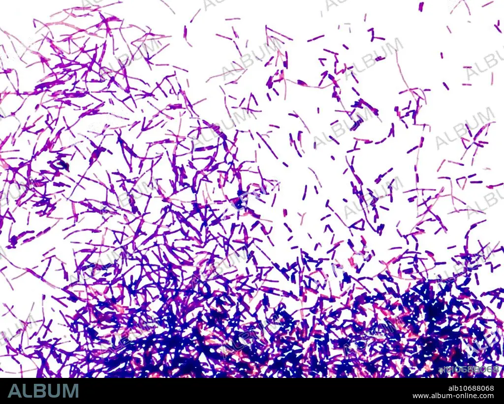
Gram-positive bacteria, light micrograph. These are bacilli (rod-shaped) bacteria. Gram staining divides bacteria into two groups (Gram-positive or Gram-negative) based on the chemical and physiological properties of their cell walls. Gram-positive bacteria retain the crystal violet dye during the Gram stain process and so appear blue or violet under a microscope. Gram-negative bacteria appear red or pink. Magnification: x2800 when printed at 10 centimetres wide.
Credit:
Album / Science Source / RICHARD J. GREEN
Releases:
Model: No - Property: No
Rights questions?
Rights questions?
Image size:
3417 x 2563 px | 25.1 MB
Print size:
28.9 x 21.7 cm | 11.4 x 8.5 in (300 dpi)
Keywords:
ANALYSING • BACILLI • BACILLUS • BACTERIA • BACTERIOLOGY • BACTERIUM • BIOLOGICAL • BIOLOGY • BLUE • BLUE. • CRYSTAL • GRAM • GRAM-POSITIVE • GRAMME • LIGHT • LM • MANY • MICROBIOLOGICAL • MICROBIOLOGY • MICROGRAPH • MICROSCOPE • MICROSCOPIC • MICROSCOPY • MULTIPLE • POSITIVE ORGAN • POSITIVE • PURPLE • SCIENCE: MICROSCOPE • STAIN • STAINED • STAINING • TESTING • VIOLET
