alb5407271
Human Abdominal Aponeurosis, Anterior,1844
| Share |
|---|
 Pinterest Pinterest |
 Twitter Twitter |
 Facebook Facebook |
 Copy link Copy link |
 Email Email |
|
Add to another lightbox |
|
Add to another lightbox |


Buy this image.
Select the use:

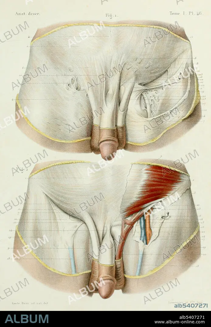
Title: Human Abdominal Aponeurosis, Anterior,1844
Caption: Plate 46. Abdominal aponeurosis. Volume 1; Osteology, syndesmology, myology of Atlas d'anatomie descriptive du corps humain by Louis Constantin Bonamy and Paul Broca with illustrations by Emile Beau, 1844. An aponeurosis is a type or a variant of the deep fascia, in the form of a sheet of pearly-white fibrous tissue that attaches sheet-like muscles needing a wide area of attachment. Their primary function is to join muscles and the body parts they act upon, whether it be bone or other muscles. The anterior abdominal aponeuroses are located just superficial to the rectus abdominis muscle. It has for its borders the external oblique, pectoralis muscles, and the latissimus dorsi. The groin is the junctional area between the abdomen and the thigh on either side of the pubic bone.
Personalities: EMILE BEAU
Credit: Album / Science Source
Releases: ? Model Release: No - ? Property Release: No
Rights questions?
Rights questions?
Image size: 3254 × 4800 px | 44.7 MB
Print size: 27.6 × 40.6 cm | 1281.1 × 1889.8 in (300 dpi)
Keywords: 1844 • 19TH CENTURY • ABDOMEN • ANATOMY • ANTERIOR • ANTERIORLY • APONEUROSES • APONEUROSIS • ATLAS D'ANATOMIE DESCRIPTIVE DU CORPS HUMAIN • BELLY • DEEP FASCIA • EMILE BEAU • EXTERNAL OBLIQUE • FIBROUS TISSUE • GROIN • GROSS ANATOMY • HEALTHY • HISTORY • HUMAN • HUMANE • INDIVIDUAL • INGUINAL REGION • LOUIS CONSTANTIN BONAMY • MALE • MUSCLE • MUSCULOSKELETAL SYSTEM • MYOLOGY • NORMAL • OSTEOLOGY • PAUL BROCA • PENE • PENILE • PENIS • PERSON • STOMACH • VOLUME 1
