alb3777231
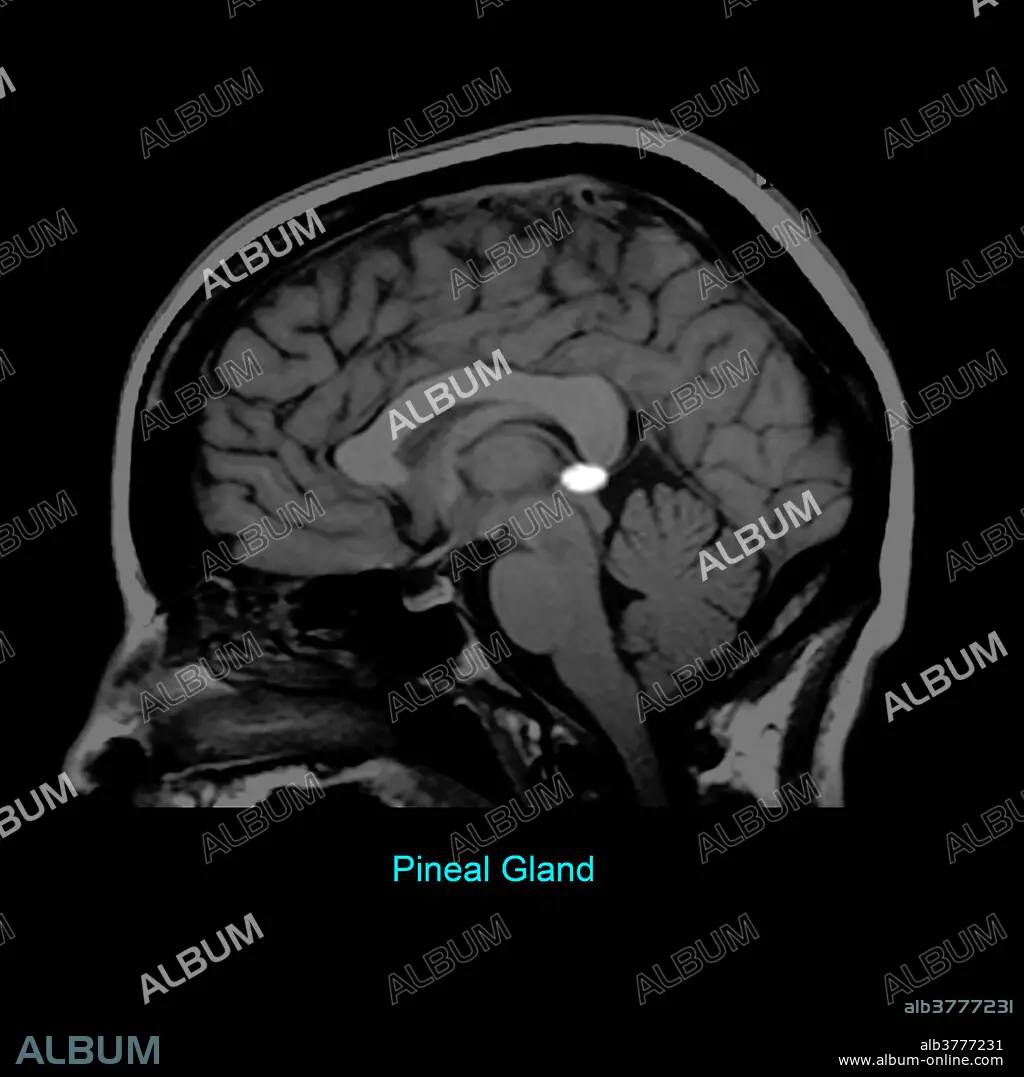
Pineal Gland, Sagittal MRI
| Share |
|---|
 Pinterest Pinterest |
 Twitter Twitter |
 Facebook Facebook |
 Copy link Copy link |
 Email Email |
|
Add to another lightbox |
|
Add to another lightbox |


Buy this image.
Select the use:

Title: Pineal Gland, Sagittal MRI
Caption: This T1 weighted sagittal (from the side) image of the brain highlights the pineal gland, which is part of the epithalamus.
Credit: Album / Science Source / Living Art Enterprises
Releases: ? Model Release: No - ? Property Release: No
Rights questions?
Rights questions?
Image size: 3600 × 3600 px | 37.1 MB
Print size: 30.5 × 30.5 cm | 1417.3 × 1417.3 in (300 dpi)
Keywords: ANATOMY • BRAIN • CEREBRAL • CEREBRI • DIAGNOSTIC • DIENCEPHALON. • EPITHALAMUS • GLAND • GROSS ANATOMY • HEALTHY • IMAGE • IMAGING • LABEL • LABELLED • LABELS • MAGNETIC • MEDICAL • MEDICINAL • MRI • NORMAL • PINEAL • RADIOGRAPHY • RADIOLOGY • REGION • RESONANCE • SCAN • SCIENCE • SONORITY • T1 • T1-WEIGHTED • WEIGHTED • WITH
