alb10697460
Egg Cell in Hamster, SEM
| Share |
|---|
 Pinterest Pinterest |
 Twitter Twitter |
 Facebook Facebook |
 Copy link Copy link |
 Email Email |
|
Add to another lightbox |
|
Add to another lightbox |


Buy this image.
Select the use:

Title:
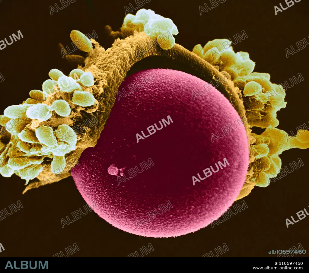
Egg Cell in Hamster, SEM
Caption:
Colour enhanced Scanning Electron Micrograph (SEM) of a hamster egg cell, showing some of the Zona pellucida and cumulus cells, most of which were removed mechanically. Magnification: 2,000X at 8x10 image size.
Credit:
Album / Science Source / David M. Phillips
Releases:
Model: No - Property: No
Rights questions?
Rights questions?
Image size:
4584 x 3812 px | 50.0 MB
Print size:
38.8 x 32.3 cm | 15.3 x 12.7 in (300 dpi)
Keywords:
ANATOMICAL • ANATOMY • BIOLOGY • CELL • CELLULAR • CUMULUS • DISC • DISCUS • DISK • EGG • ELECTRON • EM • FEMALE • FOLLICULAR • GAMETOPHYTE • GERM • GRANULOSA • GROSS ANATOMY • HAMSTER • HISTOLOGY • IMAGING • MEDICAL • MEDICINAL • MICROBIOLOGY • MICROGRAPH • MICROGRAPHY • MICROSCOPY • OOCYTE • OOPHORUS • OVOCYTE • OVUM • PELLUCIDA • PRLIGERUS • REPRODUCTIVE • SCANNING • SCIENCE • SEM • SYSTEM • ZONA
