alb10652681
Abdominal aorta, calcification, CT angiography
| Share |
|---|
 Pinterest Pinterest |
 Twitter Twitter |
 Facebook Facebook |
 Copy link Copy link |
 Email Email |
|
Add to another lightbox |
|
Add to another lightbox |


Buy this image.
Select the use:

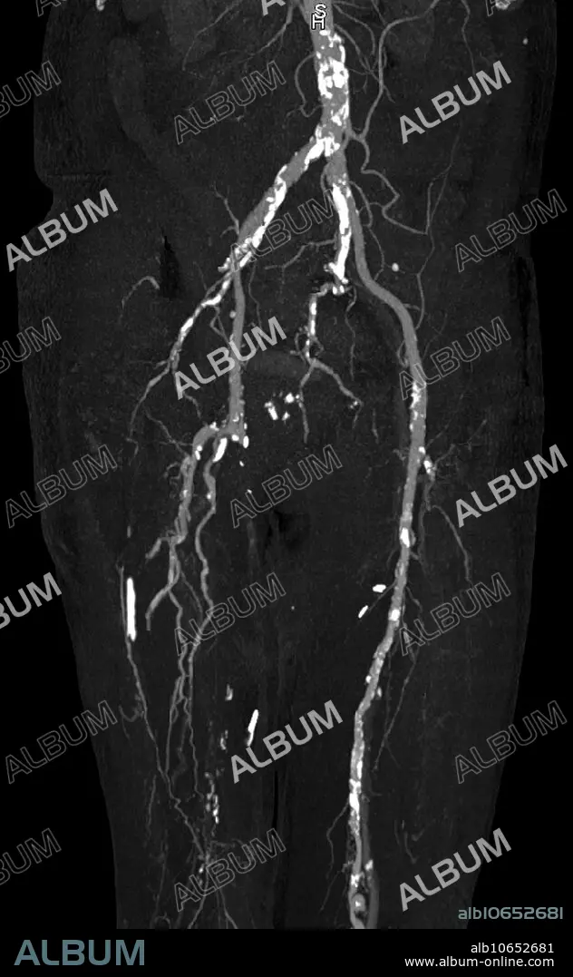
Title: Abdominal aorta, calcification, CT angiography
Caption: CT angiography of the lower extremities reveals dense calcification (bright white plaques) of abdominal aorta, femoral and popliteal arteries in a 68 year old dialysis patient with chronic renal failure and secondary hyperparathyroidism. The right femoral and popliteal arteries are occluded and collateral vessels are visible in the distal thigh.
Credit: Album / Science Source / Steven Needell
Releases: ? Model Release: No - ? Property Release: No
Rights questions?
Rights questions?
Image size: 2337 × 3787 px | 25.3 MB
Print size: 19.8 × 32.1 cm | 920.1 × 1490.9 in (300 dpi)
Keywords: ABNORMAL • ANGIOGRAM • ANGIOGRAPHY • AORTA • ARTERIA • ARTERY • ATHEROSCLEROSIS • CALCIFICATION • CLOG • CLOGGED • CLOT • COAGULUM • CONDITION • CT • DIAGNOSTIC • DISEASE • DISORDER • FAILURE • FEMORAL • FEMORIS • HYPERPARATHYROIDISM • IMAGING • MEDICAL • MEDICINAL • MEDICINE • MESS • MESSY • OCCLUSION • PATHOLOGICAL • PATHOLOGY • PLAQUE • PLATE • POPLITEAL • RADIOGRAPHY • RADIOLOGY • RENAL • SCAN • UNHEALTHY • VEIN • VENA • X-RAY
