alb9202032
Illustration of Eye Anatomy
| Share |
|---|
 Pinterest Pinterest |
 Twitter Twitter |
 Facebook Facebook |
 Copy link Copy link |
 Email Email |
|
Add to another lightbox |
|
Add to another lightbox |


Buy this image.
Select the use:

Title:
Illustration of Eye Anatomy
Caption:
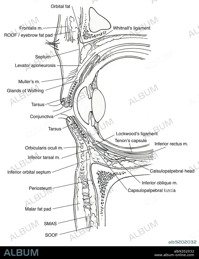
Anatomical illustration of eye, showing, orbital fat, Whitnall's ligament, Tenon's capsule, superior rectus m., levator m., inferior rectus m., Tenon's capsule, calsulopalpebral head, inferior oblique m., capsulopalpebral fascia, SOOF, SMAS, malar fat pad, periosteum, inferior orbital septum, inferior tarsal m., Lockwood's ligament, orbicularis oculi m., tarsus, conjunctiva, glands of Wolfring, Muller's m., levator aponeurosis, septum, orbicularis m., ROOF/eyebrow fat pad, and frontalis m.
Credit:
Album / Science Source
Releases:
Model: No - Property: No
Rights questions?
Rights questions?
Image size:
Not available
Print size:
Not available
Keywords:
ANATOMY • APONEUROSIS • ART • BODY • BT2987 • BW • CALSULOPALPEBRAL • CAPSULE • CAPSULOPALPEBRAL • CONJUNCTIVA • EYE • EYEBALL • EYEBALLS • EYES • FASCIA • FAT • FRONTALIS • GLANDS • GROSS ANATOMY • HEAD • HEALTHY • HUMAN • HUMANE • ILLUSTRATION • ILLUSTRATIONS • INDIVIDUAL • INFERIOR • LEVATOR • LIGAMENT • LIGAMENTUM • LOCKWOOD'S • M • MALAR • MULLER'S • NORMAL • OBLIQUE • OCULI • ORBICULARIS • ORBITAL • ORBITING • PAD • PERIOSTEUM • PERSON • RECTUS • RF • ROOF / EYEBROW • SEPTUM • SMAS • SOOF • STRUCTURE • SUPERIOR • TARSAL • TARSUS • TENON'S • WHITNALL'S • WOLFRING
