alb13913848
Rat testis, light micrograph
| Compartir |
|---|
 Pinterest Pinterest |
 Twitter Twitter |
 Facebook Facebook |
 Copiar enlace Copiar enlace |
 Email Email |
|
Añadir a otro lightbox |
|
Añadir a otro lightbox |


¿Ya tienes cuenta? Iniciar sesión
¿No tienes cuenta? Regístrate
Compra esta imagen

Título:
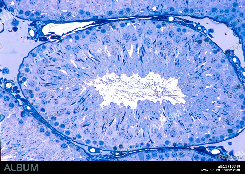
Rat testis, light micrograph
Descripción:
Traducción automática: Testículo de rata, micrografía óptica. Corte transversal de un túbulo seminífero, revestido por un epitelio seminífero donde se ubican espermatogonias y células de Sertoli en la base. Los espermatocitos primarios en el paquiteno de la profase I de la primera división meiótica ocupan una posición intermedia, y las espermátidas con núcleos alargados se encuentran cerca del lumen. Corte de 0,5 micrómetros de espesor de material plástico incrustado, teñido con azul de toluidina.
Rat testis, light micrograph. Cross-section of a seminiferous tubule, lined by a seminiferous epithelium where spermatogonia and Sertoli cells are located at the base, primary spermatocytes in pachytene of prophase I of the first meiotic division occupy an intermediate position, and spermatids with elongated nuclei are found near the lumen. 0.5 micrometre thick section of plastic embedded material stained with toluidine blue.
Crédito:
Album / JOSE CALVO / SCIENCE PHOTO LIBRARY
Autorizaciones:
Modelo: No - Propiedad: No
¿Preguntas relacionadas con los derechos?
¿Preguntas relacionadas con los derechos?
Tamaño imagen:
4800 x 3168 px | 43.5 MB
Tamaño impresión:
40.6 x 26.8 cm | 16.0 x 10.6 in (300 dpi)
Palabras clave:
BIOLOGIA • BIOLÓGICA • LUZ • MEIOSIS • MICROGRAFIA • MICROSCOPIA • NADIE • NORMALES • SALUDABLE • TESTICULOS
