alb10656341
Meiosis, light micrograph
| Compartir |
|---|
 Pinterest Pinterest |
 Twitter Twitter |
 Facebook Facebook |
 Copiar enlace Copiar enlace |
 Email Email |
|
Añadir a otro lightbox |
|
Añadir a otro lightbox |


¿Ya tienes cuenta? Iniciar sesión
¿No tienes cuenta? Regístrate
Compra esta imagen.
Selecciona el uso:

Título:
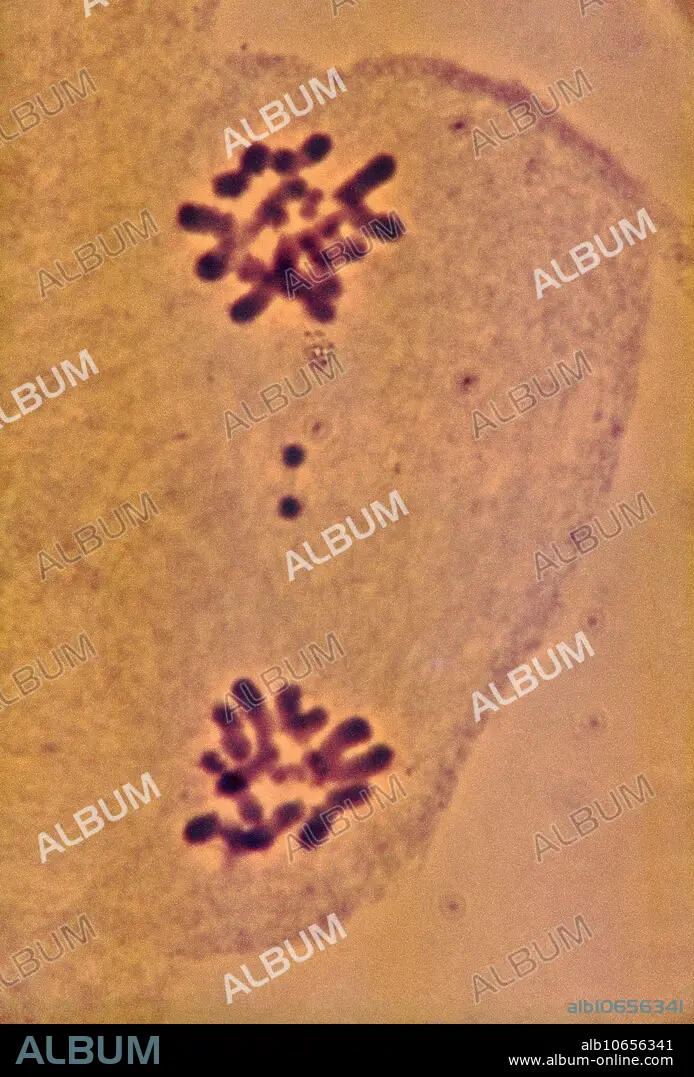
Meiosis, light micrograph
Descripción:
Ver traducción automática
Meiosis. Light micrograph of a locust (Locus testis) cell during telophase (I) of meiosis (gamete formation). During meiosis four daughter nuclei are formed from one parent nucleus after two stages of nuclear division. Meiosis occurs only in the sex cells (gametes) of the testes and ovaries. At telophase (I) pairs of homologous chromosomes have been separated and pulled to opposite poles of the cell by spindles (not seen). New nuclear membranes form around each set of chromosomes, resulting in two cells with half the usual number of chromosomes. The full complement is restored when two gametes fuse during fertilisation. Magnification: x1,500 when printed at 10 centimetres tall.
Crédito:
Album / Science Source / BIOPHOTO ASSOCIATES
Autorizaciones:
Modelo: No - Propiedad: No
¿Preguntas relacionadas con los derechos?
¿Preguntas relacionadas con los derechos?
Tamaño imagen:
3474 x 5125 px | 50.9 MB
Tamaño impresión:
29.4 x 43.4 cm | 11.6 x 17.1 in (300 dpi)
Palabras clave:
ACIDO • ADN • ANIMAL • APARATO REPRODUCTOR • BIOLOGIA • BIOLÓGICA • CELULA • CITOLOGÍA • CONDENSADA • CRECIMIENTO • CROMOSOMAS • DESOXIRRIBONUCLEICO • DIVISION • GAMETO • HAPLOIDES • LUZ • MEIOSIS • MICROGRAFIA • MICROSCOPIO • NUCLEAR • NUCLEO • REPRODUCCION • REPRODUCTION • SECUENCIA • SERIE • SEXO • TELOFASE
