alb10693434
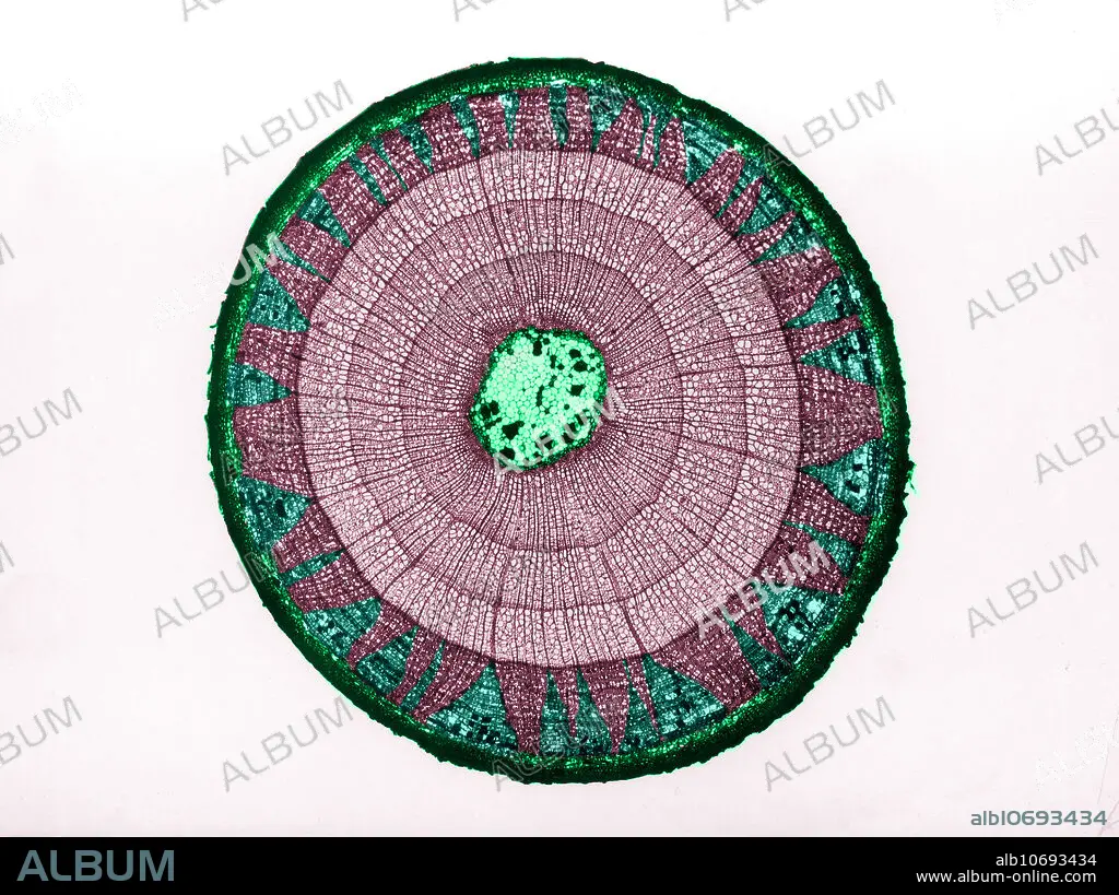
Tilia stem, LM
| Compartir |
|---|
 Pinterest Pinterest |
 Twitter Twitter |
 Facebook Facebook |
 Copiar enlace Copiar enlace |
 Email Email |
|
Añadir a otro lightbox |
|
Añadir a otro lightbox |


¿Ya tienes cuenta? Iniciar sesión
¿No tienes cuenta? Regístrate
Compra esta imagen.
Selecciona el uso:

Título: Tilia stem, LM
Descripción: Ver traducción automática
Colour enhanced light micrograph of the cross section of a Tilia sp. stem. The stem is three-years-old as it has three growth rings (concentric circles). The following is depicted (from outer to inner circle): epidermis (dark green edge), primary and secondary phloem (dark maroon section) with pith rays (green triangular parts in phloem), primary and secondary Xylem (lighter sets of rings), and Pith (green portion in centre).
Colour enhanced light micrograph of the cross section of a Tilia sp. stem. The stem is three-years-old as it has three growth rings (concentric circles). The following is depicted (from outer to inner circle): epidermis (dark green edge), primary and secondary phloem (dark maroon section) with pith rays (green triangular parts in phloem), primary and secondary Xylem (lighter sets of rings), and Pith (green portion in centre).
Crédito: Album / Science Source / Omikron
Autorizaciones: ? Cesión de modelo: No - ? Cesión de propiedad: No
¿Preguntas relacionadas con los derechos?
¿Preguntas relacionadas con los derechos?
Tamaño imagen: 4217 × 3154 px | 38.1 MB
Tamaño impresión: 35.7 × 26.7 cm | 1660.2 × 1241.7 in (300 dpi)
Palabras clave: ANATOMIA • ANGIOSPERMA • ANILLO • ANILLOS • BIOLOGIA • BOTANICA • BOTANICO • CELULA • CELULARES • CIENCIA • COLOREADA • CRECIMIENTO • DE FIBRA • ESTRUCTURA • FIBRAS • FLORA • LIBER • LUZ • MAYOR • MEDULA • MEJORA • MICROBIOLOGIA • MICROGRAFIA • NADIE • PARÉNQUIMA • PLACAS • PLANTA • PLATO • PRIMARIA • TALLO • TILIA • XILEMA
