alb10671444
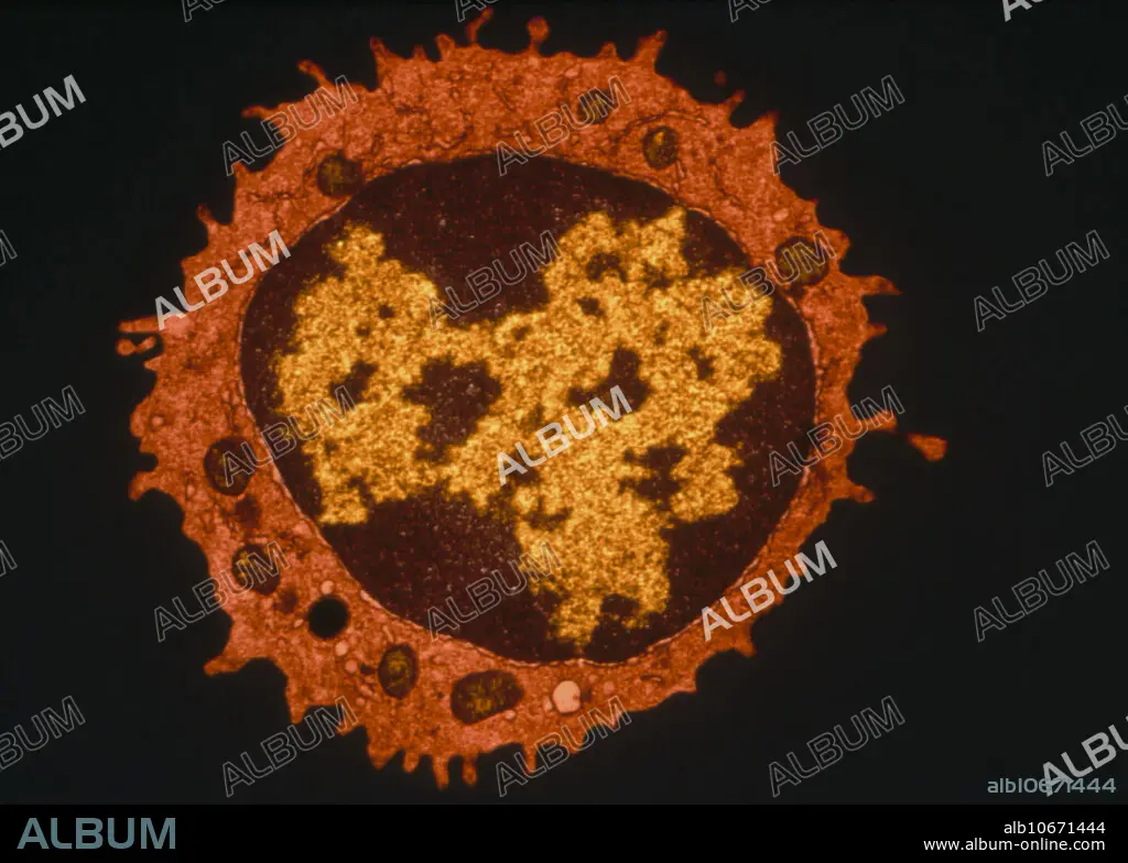
Coloured TEM of a T-lymphocyte white blood cell
| Compartir |
|---|
 Pinterest Pinterest |
 Twitter Twitter |
 Facebook Facebook |
 Copiar enlace Copiar enlace |
 Email Email |
|
Añadir a otro lightbox |
|
Añadir a otro lightbox |


¿Ya tienes cuenta? Iniciar sesión
¿No tienes cuenta? Regístrate
Compra esta imagen.
Selecciona el uso:

Título:
Coloured TEM of a T-lymphocyte white blood cell
Descripción:
Ver traducción automática
T-lymphocyte. Coloured transmission electron micrograph (TEM) of a section through a T-lympho- cyte white blood cell. At centre, the large nucleus is seen (brown) with chromatin (yellow). Characteristic of normal T-lymphocytes are long microvilli which project from the cell surface. Mitochondria (brown) are seen in the cell cytoplasm (orange). T-lymphocytes are cells of the human immune system, produced in bone marrow and which mature in the thymus gland. They help to protect the body against invasion by bacteria, viruses and other foreign substances. T-cells are attacked by the human immunodeficiency virus (HIV) which causes AIDS.
Crédito:
Album / Science Source / DON FAWCETT
Autorizaciones:
Modelo: No - Propiedad: No
¿Preguntas relacionadas con los derechos?
¿Preguntas relacionadas con los derechos?
Tamaño imagen:
3543 x 2528 px | 25.6 MB
Tamaño impresión:
30.0 x 21.4 cm | 11.8 x 8.4 in (300 dpi)
