alb10660588
Tongue cancer, PET CT scan
| Compartir |
|---|
 Pinterest Pinterest |
 Twitter Twitter |
 Facebook Facebook |
 Copiar enlace Copiar enlace |
 Email Email |
|
Añadir a otro lightbox |
|
Añadir a otro lightbox |


¿Ya tienes cuenta? Iniciar sesión
¿No tienes cuenta? Regístrate
Compra esta imagen.
Selecciona el uso:

Título:
Tongue cancer, PET CT scan
Descripción:
Ver traducción automática
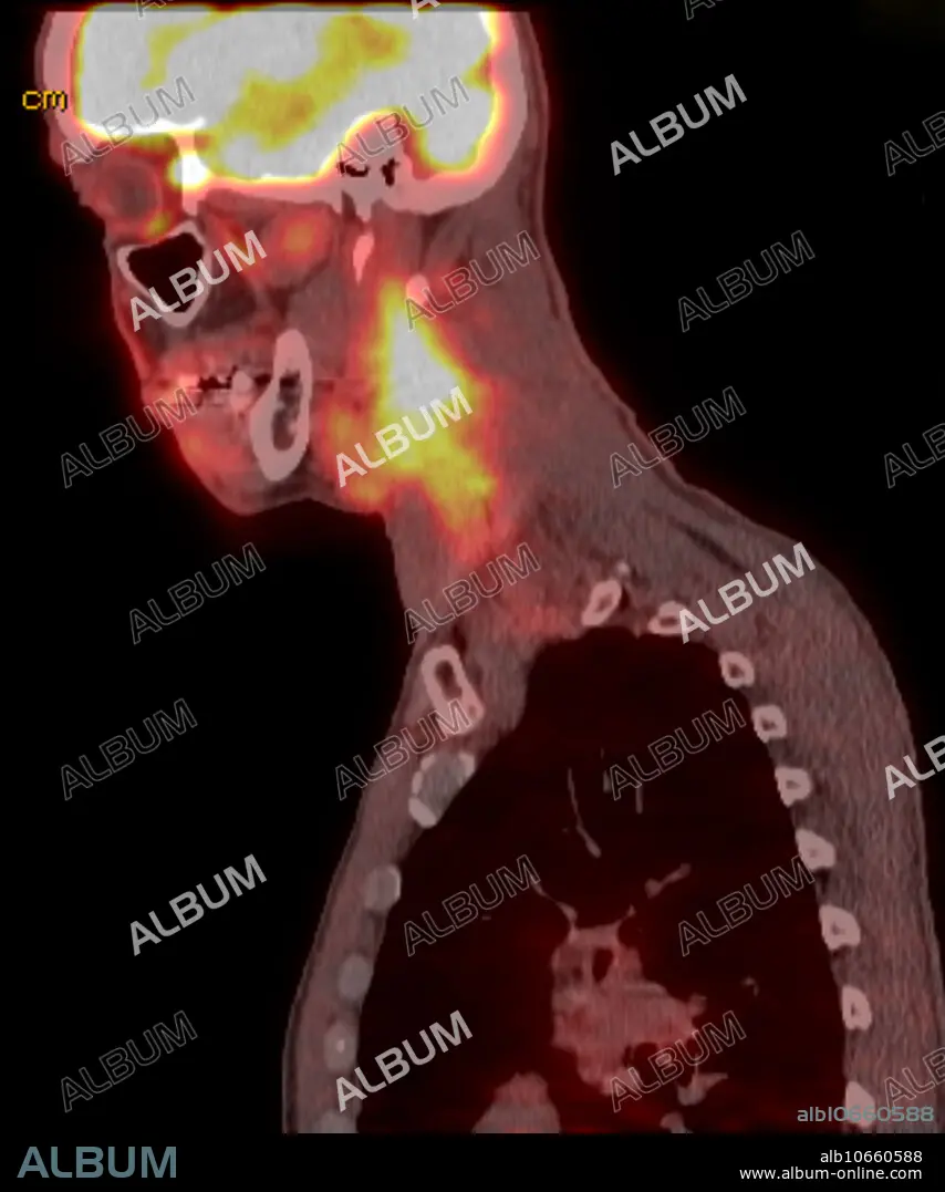
Positron emission tomography of 60 yo male with right squamous cell carcinoma of the tongue. Sagittal FDG CT PET scan reveals large conglomeration of lymph nodes within the right neck beginning at the angle of the mandible and extending to the level lower neck (level 5) and also extension into the posterior triangle. The mass is deep to the sternocleidomastoid. Mass extends posteriorly into the posterior triangle. Corresponding intense metabolic uptake with SUV of 6.0. There is focal asymmetric uptake involving the base the tongue, with SUV of 4.9.
Personas:
Crédito:
Album / Science Source / Steven Needell
Autorizaciones:
Tamaño imagen:
1454 x 1744 px | 7.3 MB
Tamaño impresión:
12.3 x 14.8 cm | 4.8 x 5.8 in (300 dpi)
Palabras clave:
60 • ANORMAL • ARTERIA • CANCER • CARCINOMA • CAROTIDA • CELULA • CONDICION • CUELLO • DESORDEN • EMISION • ENFERMEDAD • ESCAMOSAS • ESTERNOCLEIDOMASTOIDEO • ESTERNOCLEIDOMASTOIDEOS • GATO • HEMORRAGIA • IMAGENES • INSALUBRE • MASCULINO • MASS • MEDICINA • MEDICINAL • MÚSCULO ESTERNOCLEIDOMASTOIDEO • PATOLOGIA • PATOLÓGICOS • RADIOLOGIA • SAGITAL • SANGRADURA • SANGRANDO • SANGRANTE • SANGRANTES • TIROIDES, LA • TOMOGRAFÍA • VOLUMEN
