alb3774451
Lumbar Compression Fracture, Illustration
| Compartir |
|---|
 Pinterest Pinterest |
 Twitter Twitter |
 Facebook Facebook |
 Copiar enlace Copiar enlace |
 Email Email |
|
Añadir a otro lightbox |
|
Añadir a otro lightbox |


¿Ya tienes cuenta? Iniciar sesión
¿No tienes cuenta? Regístrate
Compra esta imagen.
Selecciona el uso:

Título: Lumbar Compression Fracture, Illustration
Descripción: Ver traducción automática
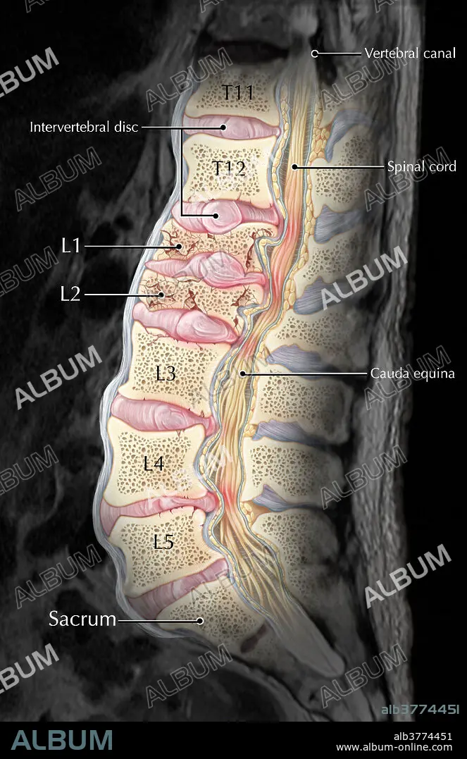
An interpretive illustration of an MRI depicting a sagittal view of compression fractures at the L1 and L2 vertebrae as a result of osteoporosis. Over time as bone becomes weaker and more porous, they become more susceptible to injury and fractures, especially in situations where significant weight or stress is placed on the bone. In this case, the vertebral bodies of L1 and L2 have collapsed, resulting in a displacement of the bones and intervertebral discs into the spinal canal, resulting in pain and possibly reducing the patient's mobility.
An interpretive illustration of an MRI depicting a sagittal view of compression fractures at the L1 and L2 vertebrae as a result of osteoporosis. Over time as bone becomes weaker and more porous, they become more susceptible to injury and fractures, especially in situations where significant weight or stress is placed on the bone. In this case, the vertebral bodies of L1 and L2 have collapsed, resulting in a displacement of the bones and intervertebral discs into the spinal canal, resulting in pain and possibly reducing the patient's mobility.
Crédito: Album / Science Source / Evan Oto
Autorizaciones: ? Cesión de modelo: No - ? Cesión de propiedad: No
¿Preguntas relacionadas con los derechos?
¿Preguntas relacionadas con los derechos?
Tamaño imagen: 3300 × 5100 px | 48.2 MB
Tamaño impresión: 27.9 × 43.2 cm | 1299.2 × 2007.9 in (300 dpi)
Palabras clave: ANORMAL • ARTE • CANAL • COMPRESION • DISCO • DISK • ENFERMEDAD • ESPINAL • FONDO • FOTO • HERNIA • HUESO • HUESOS • ILUSTRACION • ILUSTRACIONES • IMAGEN • IMAGENES • LUMBAR • MEDICINAL • NEGRO • OBRA DE ARTE • OSTEOPOROSIS • PATOLOGIA • POROSA • RESONANCIA • RM • SAGITAL • SE DESLIZÓ • TORACICA • VERTEBRALES
