alb5575977
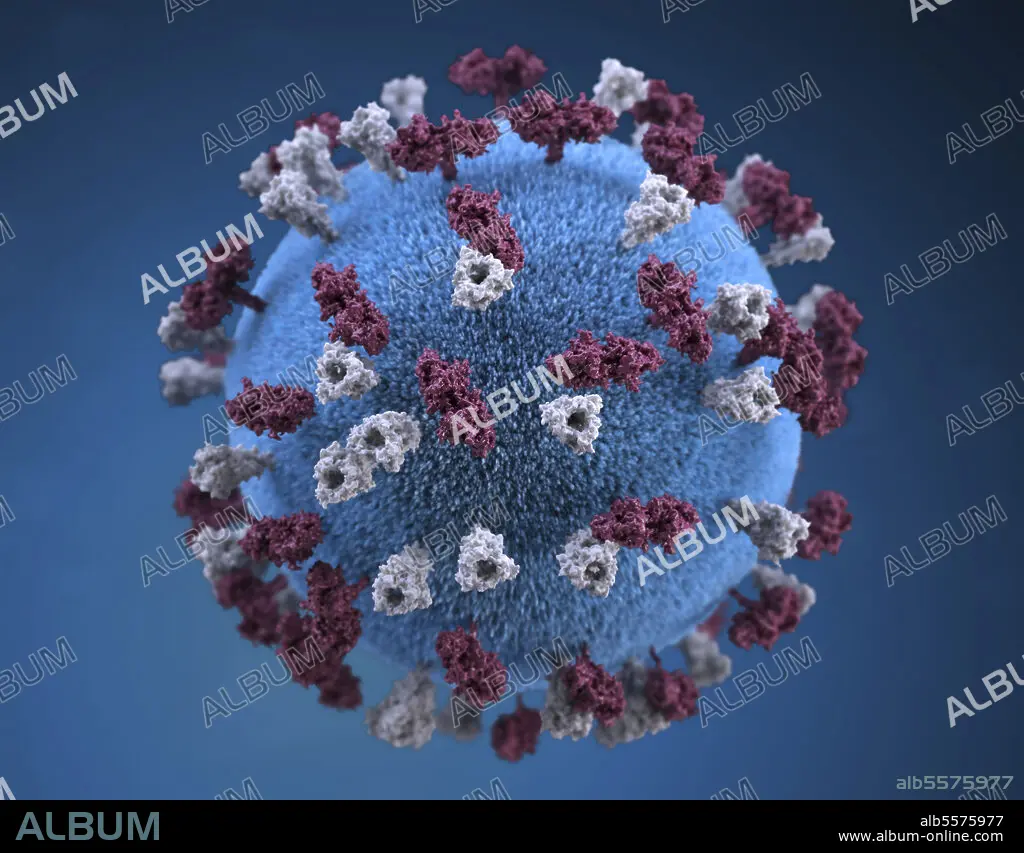
3D illustration of spherical-shaped, measles virus particle.
| Compartir |
|---|
 Pinterest Pinterest |
 Twitter Twitter |
 Facebook Facebook |
 Copiar enlace Copiar enlace |
 Email Email |
|
Añadir a otro lightbox |
|
Añadir a otro lightbox |


¿Ya tienes cuenta? Iniciar sesión
¿No tienes cuenta? Regístrate
Compra esta imagen.
Selecciona el uso:

Título: 3D illustration of spherical-shaped, measles virus particle.
Descripción: Ver traducción automática
3D illustration of spherical-shaped, measles virus particle, that is studded with glycoprotein tubercles. These tubercular studs colorized maroon, are known as H-proteins (hemagglutinin), while those colorized gray, represented what are referred to as F-proteins (fusion). The F-protein is responsible for fusion of the virus and host cell membranes, viral penetration, and hemolysis. The H-protein is responsible for the binding of virions to cells. Both types of proteinaceous studs are embedded in the particle envelopes lipid bilayer.
3D illustration of spherical-shaped, measles virus particle, that is studded with glycoprotein tubercles. These tubercular studs colorized maroon, are known as H-proteins (hemagglutinin), while those colorized gray, represented what are referred to as F-proteins (fusion). The F-protein is responsible for fusion of the virus and host cell membranes, viral penetration, and hemolysis. The H-protein is responsible for the binding of virions to cells. Both types of proteinaceous studs are embedded in the particle envelopes lipid bilayer.
Crédito: Album / Stocktrek Images
Tamaño imagen: 3600 × 2813 px | 29.0 MB
Tamaño impresión: 30.5 × 23.8 cm | 1417.3 × 1107.5 in (300 dpi)
Palabras clave: ÁCIDO RIBONUCLEICO • ANATOMIA • AUMENTO • AZUL • BACTERIAS • BIOLOGÍA CELULAR • BIOLOGIA MOLECULAR • BIOLOGIA • BIOMÉDICA • BIOQUIMICA • CELULA • CIENCIA • CITOLOGÍA • CITOPLASMA • CONTAGIOSO • EN TRES DIMENSIONES • ENDÉMICAS • ENFERMEDAD • ENFERMEDADES INFECCIOSAS • EPIDEMIOLOGÍA • HORIZONTAL • ILUSTRACION • ILUSTRACIONES BIOMÉDICAS • ILUSTRACIONES • IMAGEN EN COLOR • IMAGEN GENERADA DIGITALMENTE • INFECCION • INFECCIOSAS • INVESTIGACION • LÍPIDOS • MEDICINA • MEDICINAL • MICROBIO • MICROBIOLOGIA • MICROGRAFIA • MICROSCOPIA • MICROSCÓPICO • MICROSCOPIO ELECTRÓNICO DE BARRIDO • ORGANISMO • PANDEMIA • PATOLOGIA • PRIMER PLANO • PROTEINAS • PROTOPLASMA • RNA • SALUD • SARAMPIÓN • SIN GENTE • SOLO OBJETO • SUPERFICIE • TUBERCULO • ULTRAESTRUCTURA • VIROLOGIA • VIRUS
