alb10690951
Intestinal microvilli, TEM
| Compartir |
|---|
 Pinterest Pinterest |
 Twitter Twitter |
 Facebook Facebook |
 Copiar enlace Copiar enlace |
 Email Email |
|
Añadir a otro lightbox |
|
Añadir a otro lightbox |


¿Ya tienes cuenta? Iniciar sesión
¿No tienes cuenta? Regístrate
Compra esta imagen.
Selecciona el uso:

Título:
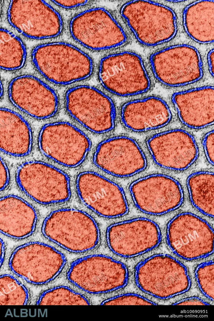
Intestinal microvilli, TEM
Descripción:
Ver traducción automática
Transmission electron micrograph of the intestinal microvilli of a cat. The cross-section of each microvillus (in red) is bounded by two dense lines of similar thickness (in black) separated by a lighter intermediate zone (in blue). Not all cell membranes exhibit his degree of symmetry. In some, the outer dense line is thinner than the inner.
Crédito:
Album / Science Source / Don W. Fawcett
Autorizaciones:
Modelo: No - Propiedad: No
¿Preguntas relacionadas con los derechos?
¿Preguntas relacionadas con los derechos?
Tamaño imagen:
3489 x 4962 px | 49.5 MB
Tamaño impresión:
29.5 x 42.0 cm | 11.6 x 16.5 in (300 dpi)
Palabras clave:
ABSORCIÓN • CELULA • COLOREADA • ELECTRON • EPITELIO • FRONTERA • INTESTINALES • MAMÍFEROS • MEJORA • MEJORAR • MEMBRANA • MICROGRAFIA • MICROSCOPIA • NUTRIENTES • SISTEMA • TEMPERATURA • TRANSMISION
