alb3808882
Gangrene, Illustration, 1830s
| Compartir |
|---|
 Pinterest Pinterest |
 Twitter Twitter |
 Facebook Facebook |
 Copiar enlace Copiar enlace |
 Email Email |
|
Añadir a otro lightbox |
|
Añadir a otro lightbox |


¿Ya tienes cuenta? Iniciar sesión
¿No tienes cuenta? Regístrate
Compra esta imagen.
Selecciona el uso:

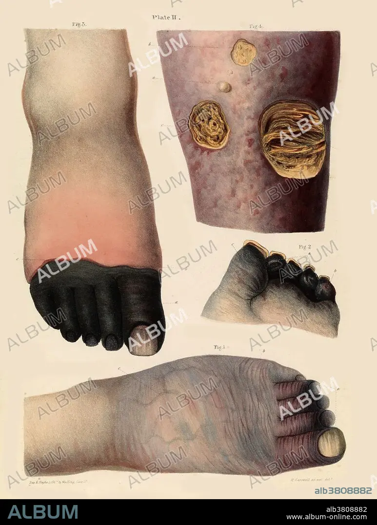
Título: Gangrene, Illustration, 1830s
Descripción:
Traducción automática: Ilustración de gangrena de la década de 1830. Las figuras 1 y 2 (abajo y centro derecha) muestran gangrena senilis: decoloración de los dedos de los pies al comienzo de la enfermedad. La figura 3 (arriba a la izquierda) muestra la gangrena de los dedos de los pies. La figura 4 (arriba a la derecha) muestra la mortificación de la piel y del tejido celular subyacente debido a un obstáculo al retorno de la sangre venosa como consecuencia de una enfermedad del corazón. Robert Carwell
Gangrene illustration from the 1830s. Figs 1 and 2 (bottom and centre right) show gangrena senilis - discoloration of the toes at the beginning of the disease. Fig 3 (top left) shows gangrene of the toes. Fig 4 (top right) shows mortification of the skin and subjacent cellular tissue from an obstacle to the return of the venous blood in consequence of disease of the heart. Robert Carswell.
Crédito: Album / Science Source / Wellcome Images
Autorizaciones: ? Cesión de modelo: No - ? Cesión de propiedad: No
¿Preguntas relacionadas con los derechos?
¿Preguntas relacionadas con los derechos?
Tamaño imagen: 4952 × 6556 px | 92.9 MB
Tamaño impresión: 41.9 × 55.5 cm | 1949.6 × 2581.1 in (300 dpi)
Palabras clave: 1800S • ADVANCED • ANORMAL • ARTE • CONDICION • DECOLORACIÓN • DEDO DEL PIE • DERMATOLOGIA • DERMATOLÓGICO • DESORDEN • DIBUJO • ENFERMEDAD DE LA PIEL • ENFERMEDAD PIEL • ENFERMEDAD • FOOT • GANGRENA • GENTE • HISTORIA • HISTORICO • ILUSTRACION • ILUSTRACIONES • MEDICINA • MEDICINAL • MORTIFICACION • OBRA DE ARTE • PACIENTE • PATOLOGIA • PERSONA • PIE • PIERNA • PIERNAS • SIGLO XIX
