alb3770261
Breast Anatomy, Illustration
| Compartir |
|---|
 Pinterest Pinterest |
 Twitter Twitter |
 Facebook Facebook |
 Copiar enlace Copiar enlace |
 Email Email |
|
Añadir a otro lightbox |
|
Añadir a otro lightbox |


¿Ya tienes cuenta? Iniciar sesión
¿No tienes cuenta? Regístrate
Compra esta imagen.
Selecciona el uso:

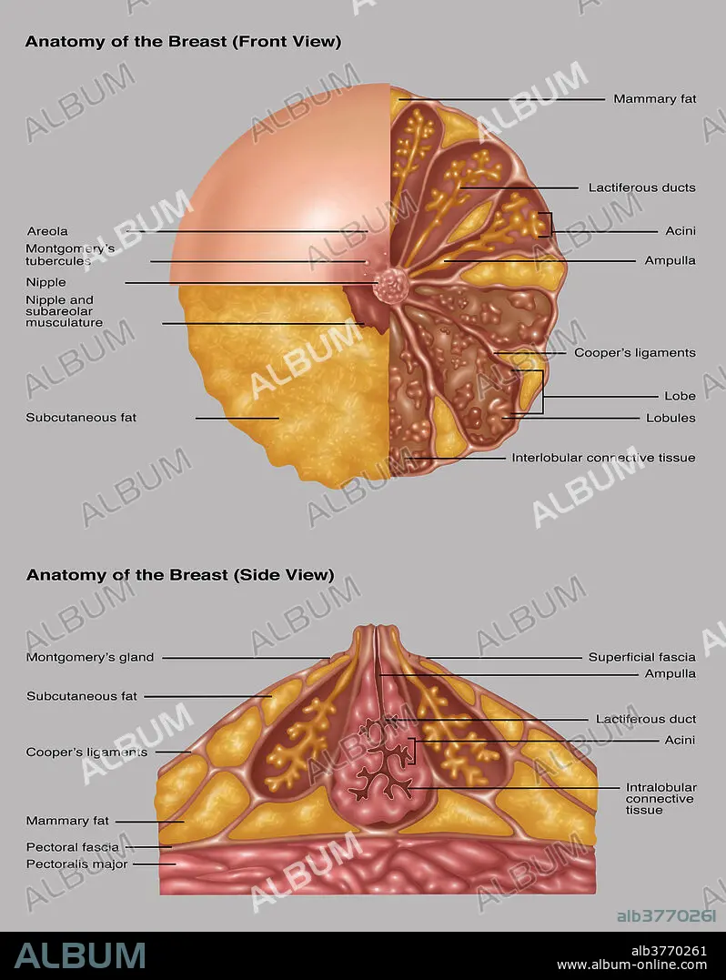
Título: Breast Anatomy, Illustration
Descripción: Ver traducción automática
Illustration detailing the anatomy of a female breast from a front view (top) and a side view (bottom). The top image is in three sections. The areola, montgomery's tubercules, and nipple are in the light pink section. The nipple and subareolar musculature and subcutaneous fat are in the orange section. On the right half is: mammary fat (orange), lactiferous ducts (orange stems), acini (end of orange stems), ampulla (on orange stem), coopers ligaments (light pink strands), lobe and lobules (brown outlines and pink groups inside the brown), and interlobular connective tissue (brown area). The bottom image shows montgomery's gland and superficial fascia (outer lining), subcutaneous fat (orange outer sections), ampulla and lactiferous duct and connective tissue (pink centre), coopers ligaments, mammary fat (orange bottom sections), pectoral fascia, and pectoralis major (pink at bottom).
Illustration detailing the anatomy of a female breast from a front view (top) and a side view (bottom). The top image is in three sections. The areola, montgomery's tubercules, and nipple are in the light pink section. The nipple and subareolar musculature and subcutaneous fat are in the orange section. On the right half is: mammary fat (orange), lactiferous ducts (orange stems), acini (end of orange stems), ampulla (on orange stem), coopers ligaments (light pink strands), lobe and lobules (brown outlines and pink groups inside the brown), and interlobular connective tissue (brown area). The bottom image shows montgomery's gland and superficial fascia (outer lining), subcutaneous fat (orange outer sections), ampulla and lactiferous duct and connective tissue (pink centre), coopers ligaments, mammary fat (orange bottom sections), pectoral fascia, and pectoralis major (pink at bottom).
Crédito: Album / Science Source / Gwen Shockey
Autorizaciones: ? Cesión de modelo: No - ? Cesión de propiedad: No
¿Preguntas relacionadas con los derechos?
¿Preguntas relacionadas con los derechos?
Tamaño imagen: 4824 × 6192 px | 85.5 MB
Tamaño impresión: 40.8 × 52.4 cm | 1899.2 × 2437.8 in (300 dpi)
Palabras clave: ANATOMIA • ANOTADO • AREOLA • ARTE • BARRILERO • CONDUCTO • CONDUCTOS • DIAGRAMA • FASCIA • GLÁNDULA • GRAFICO • ILUSTRACION • ILUSTRACIONES • INFOGRAFÍA • LIGAMENTOS • LOBULO • MAMARIA • MEDICINAL • MUSCULATURA • OBRA DE ARTE • PECTORAL • PEZON • SUBCUTÁNEA • SUPERFICIALES • TONELERO
