alb3794138
Pineal Gland
| Compartir |
|---|
 Pinterest Pinterest |
 Twitter Twitter |
 Facebook Facebook |
 Copiar enlace Copiar enlace |
 Email Email |
|
Añadir a otro lightbox |
|
Añadir a otro lightbox |


¿Ya tienes cuenta? Iniciar sesión
¿No tienes cuenta? Regístrate
Compra esta imagen.
Selecciona el uso:

Título: Pineal Gland
Descripción: Ver traducción automática
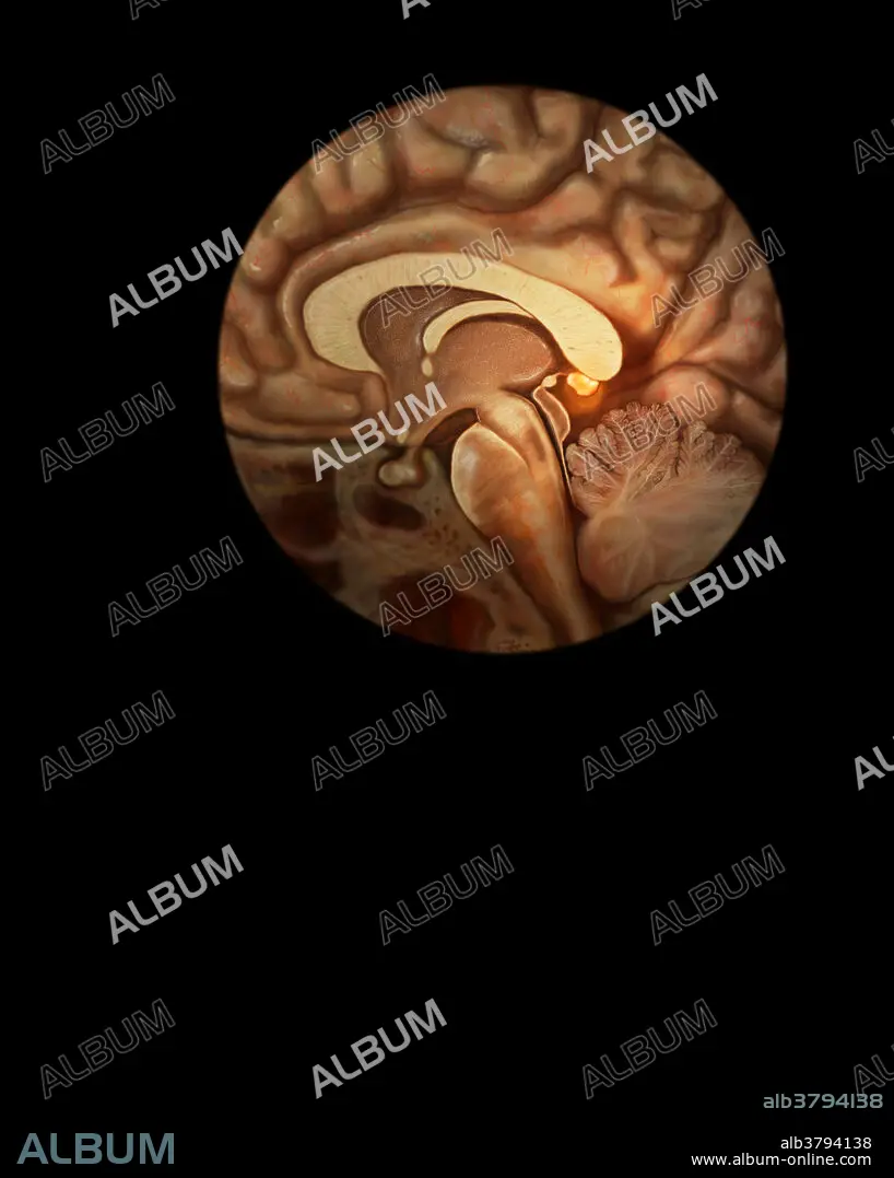
Three-dimensional visualisation based on segmented human data of the pineal gland (yellow), a small organ located on the posterior part of the roof of the third ventricle, seen here below the corpus collosum. It is connected to the brain via a short stalk containing nerve fibers which communicate with the hypothalamus. The pineal gland secretes the hormone melatonin which regulates the circadian rhythms of the body. Its secretion during hours of darkness produces a hypnotic effect which results in sleep.
Three-dimensional visualisation based on segmented human data of the pineal gland (yellow), a small organ located on the posterior part of the roof of the third ventricle, seen here below the corpus collosum. It is connected to the brain via a short stalk containing nerve fibers which communicate with the hypothalamus. The pineal gland secretes the hormone melatonin which regulates the circadian rhythms of the body. Its secretion during hours of darkness produces a hypnotic effect which results in sleep.
Crédito: Album / Science Source / ANATOMICAL TRAVELOGUE
Autorizaciones: ? Cesión de modelo: No - ? Cesión de propiedad: No
¿Preguntas relacionadas con los derechos?
¿Preguntas relacionadas con los derechos?
Tamaño imagen: 4074 × 5100 px | 59.4 MB
Tamaño impresión: 34.5 × 43.2 cm | 1603.9 × 2007.9 in (300 dpi)
Palabras clave: ANATOMIA • ANATOMICAS • CUERPO • DE FIBRA • DORMIR • DURMIENDO • FIBRAS • GLÁNDULA • GLÁNDULAS • HIPNOSIS • HIPNÓTICO • HIPOTÁLAMO • HIPOTÁLAMOS • HOMEOSTASIS • HORMONA • HUMANO • HUMANOS • ILUSTRACION • ILUSTRACIONES • MEDICINAL • NERVIO • NEURONA • PERSONA • PINEAL • SISTEMA • SISTEMAS DE, LOS • SISTEMAS • SLEEP • SOÑAR • SUEÑO
