alb10657099
Lingual Thyroid, CT Scan
| Compartir |
|---|
 Pinterest Pinterest |
 Twitter Twitter |
 Facebook Facebook |
 Copiar enlace Copiar enlace |
 Email Email |
|
Añadir a otro lightbox |
|
Añadir a otro lightbox |


¿Ya tienes cuenta? Iniciar sesión
¿No tienes cuenta? Regístrate
Compra esta imagen.
Selecciona el uso:

Título:
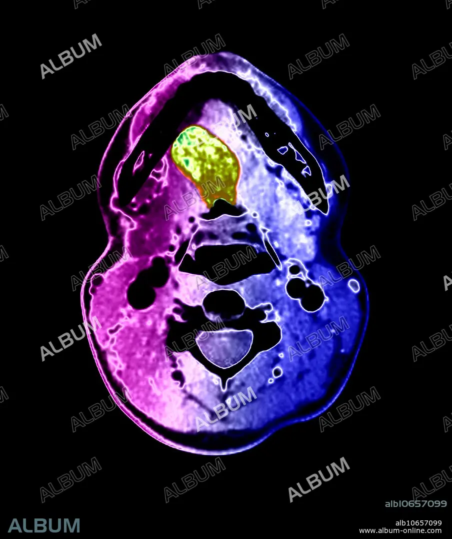
Lingual Thyroid, CT Scan
Descripción:
Ver traducción automática
This colour-enhanced, axial (cross-sectional) CT image through the oral cavity demonstrates a large, abnormal density (green) in the tongue. This represents lingual thyroid tissue. This patient had no thyroid tissue in the expected region of the lower neck. The thyroid gland develops near the foramen cecum of the tongue and normally descends along a tract called the thyroglossal duct to the lower neck. Rarely, this normal descent does not occur and thyroid tissue remains in the tongue, but remnants of thyroid tissue and the duct itself may occur anywhere along the tract. Often, however, no thyroid tissue is present in the lower neck, only in the tongue base, as in this case.
Personas:
Crédito:
Album / Science Source / Living Art Enterprises
Autorizaciones:
Modelo: No - Propiedad: No
¿Preguntas relacionadas con los derechos?
¿Preguntas relacionadas con los derechos?
Tamaño imagen:
4200 x 4755 px | 57.1 MB
Tamaño impresión:
35.6 x 40.3 cm | 14.0 x 15.8 in (300 dpi)
